
Uterin İnertiye Bağlı Distosili Kedilerde Serum Glukoz ve Kalsiyum Konsantrasyonları: Retrospektif Bir Analiz
Volume 40, Issue 3
October 2026 · Pages 216-221
Uterin İnertiye Bağlı Distosili Kedilerde Serum Glukoz ve Kalsiyum Konsantrasyonları: Retrospektif Bir Analiz
Serum Glucose and Calcium Concentrations in Cats with Dystocia due to Uterine Inertia: A Retrospective Analysis
Mehmet YILDIZ1, Sait ŞENDAĞ1, Doreen HARDEGEN2, Axel WEHREND2
2Justus-Liebig University, Veterinary Clinic for Reproductive Medicine and Neonatology, Giessen, GERMANY
Bu retrospektif çalışma, uterus tembelliği tanısı konulan güç doğumlu (distosili) dişi kedilerde serum glukoz ve iyonize kalsiyum (iCa) konsantrasyonlarını analiz etmeyi ve bu metabolik parametrelerin kedi distosisinin patogenezindeki rolüne ilişkin literatürdeki mevcut boşluğu gidermeyi amaçlamıştır. Çalışmaya farklı ırk ve yaşlardan, ortalama gebelik süresi 65±5.4 gün olan toplam 30 dişi kedi dahil edilmiştir. Ortalama serum glukoz ve iCa konsantrasyonları sırasıyla 6.96±1.94 mmol/L ve 1.31±0.20 mmol/L olarak belirlenmiştir. Serum glukoz düzeyleri, 5.00 mmol/L referans ortalama değere kıyasla istatistiksel olarak anlamlı derecede yüksek bulunmuş (p<0.01) ve olguların %43.3’ünde hiperglisemi saptanmıştır. Buna karşın, serum iCa konsantrasyonları ile 1.26 mmol/L referans değeri arasında istatistiksel olarak anlamlı bir fark bulunmamıştır (p=0.09). Hipoglisemi yalnızca bir olguda (%3.3), hipokalsemi ise altı olguda (%20) gözlenmiştir. Ayrıca, serum glukoz ve iCa düzeyleri arasında zayıf ve istatistiksel olarak anlamlı olmayan negatif bir korelasyon belirlenmiştir (r=-0.08, p=0.671). Elde edilen bulgular, uterin inertia ile ilişkili distosinin patogenezinde hipokalsemi ve hipogliseminin primer bir rol oynamadığını, gözlenen hipergliseminin ise muhtemelen doğum sürecine bağlı stresin bir sonucu olduğunu düşündürmektedir. Bu sonuçlar, glukoz ve kalsiyum düzeylerinin tek başına kedi distosisinin yönetiminde tanısal veya prognostik belirteçler olarak yeterli olmadığını göstermektedir.
This retrospective study aimed to analyze serum glucose and ionized calcium (iCa) concentrations in dystocic queens diagnosed with uterine inertia and to address the current gap in the literature regarding the role of these metabolic parameters in the pathogenesis of feline dystocia. Thirty queens of various breeds and ages (mean gestation length: 65±5.4 days) presenting with dystocia were included in the analysis. The mean serum glucose and iCa concentrations were 6.96±1.94 mmol/L and 1.31±0.20 mmol/L, respectively. Serum glucose levels were significantly elevated compared to the reference mean of 5.00 mmol/L (p<0.01), with 43.3% of cases exhibiting hyperglycemia. In contrast, no statistically significant difference was observed for serum iCa concentrations compared with the reference value of 1.26 mmol/L (p=0.09). Hypoglycemia was observed in only one case (3.3%), and hypocalcemia in six cases (20%). Additionally, a weak and non-significant negative correlation was found between serum glucose and iCa levels (r=-0.08, p=0.671). Hypocalcemia and hypoglycemia do not appear to play a primary role in the pathogenesis of dystocia associated with uterine inertia in queens, and the observed hyperglycemia is likely a consequence of parturition-related stress rather than an etiological factor. These findings suggest that glucose and calcium levels alone are insufficient as diagnostic or prognostic markers for managing feline dystocia.
Introduction
Difficult labour (dystocia) in queens is a critical clinical condition associated with both maternal and fetal mortality that often requires urgent veterinary intervention and may originate from either maternal or fetal causes [1,2]. Among maternal factors, uterine contractions play a pivotal role in initiating and sustaining labour, and inadequate or absent uterine contractions, referred to as uterine inertia, are commonly implicated in maternally induced dystocia; indeed, most cases of dystocia in queens are maternal in origin, with uterine inertia representing the predominant diagnosis [3-8]. However, our current understanding of the continuity, strength, or cessation of uterine contractions in feline parturition remains largely theoretical and unverified. For instance, some queens are capable of delivering large litters without complications, while others may fail to deliver even a few kittens due to an absence of uterine contractions. Such clinical discrepancies raise the question of whether maternal blood parameters might be involved in the pathophysiology of uterine inertia. In addition to oxytocin, glucose serves as a critical energy source, and calcium is an essential ion for maintaining uterine contractility [9]. Calcium plays a central role in neuromuscular transmission and smooth muscle contraction, both of which are vital for successful parturition. Mineral imbalances particularly hypocalcemia have been implicated in labour dysfunction. A calcium deficiency may reduce the responsiveness of the myometrium to oxytocin stimulation [10]. Moreover, fetal skeletal mineralisation in late gestation increases maternal calcium demands, potentially predisposing to hypocalcemia and subsequent uterine inertia due to impaired myometrial contractility [11]. In canine species, both perinatal hypoglycemia likely caused by increased energy demands and stress- or cortisol-induced perinatal hyperglycemia have been documented [9,12-14]. However, comparable data for queens are lacking. To date, only two studies have addressed this issue in cats [8,15]. Interestingly, their findings challenge traditional assumptions. Bailin et al. [15] reported that dystocic queens were neither hypocalcemic nor hypoglycemic in a cohort of 19 animals. Conversely, Axnér et al. [8] demonstrated that calcium supplementation with calcium chloride dihydrate was effective in treating uterine inertia in a subset of queens, suggesting a role for hypocalcemia in the condition's etiology. Furthermore, they identified an association between hyperglycemia and increased pre-discharge kitten mortality. These conflicting results, combined with the limited number of available studies, underscore the need for further research into the role of blood glucose and ionized calcium (iCa) levels in feline dystocia. Therefore, the present study was designed to investigate the relationship between serum glucose and iCa concentrations and dystocia due to uterine inertia in queens, using a larger sample size.The specific objectives of this study were to: (i) determine the distribution of blood glucose and iCa levels in dystocic queens; (ii) evaluate the correlation between glucose and iCa concentrations in this population, as this has not previously been assessed; and (iii) explore the potential role of deviations from reference values in the occurrence of uterine inertia.
Material and Method
Research and Publication Ethics: This study was conducted retrospectively using clinical records. The Van Yuzuncu Yil University Animal Researches Local Ethics Committee (VAN YUHADYEK) has reviewed the project and decided that Animal Researches Ethic Committee Approval is not required (Date: 26/02/2026, Decision number: 2026/02-26)This retrospective observational study included 30 queens of various breeds (primarily European pedigree and British shorthair breeds) and ages (1–3 years) that presented to our clinic with dystocia between 2000 and 2001. The study population consisted of both primiparous queens and queens that had previously delivered one or more litters (multiparous queens). Data were retrieved from the clinic’s electronic documentation system using the keyword “queen dystocia.” None of the queens had received any obstetric intervention prior to presentation. The mean gestation length, calculated from the date of breeding, was 65±5.4 days (range: 59.6–70.4 days). Inclusion criteria consisted of stage 2 labour and a diagnosis of dystocia based on the criteria described by Bailin et al. [15] and Sendag et al. [2]. Based on the anamnestic information, the presence of active labour, abdominal straining, and, in several cases, the passage of fetal membranes or amniotic fluids, most cases were considered clinically consistent with secondary uterine inertia. However, due to the retrospective nature of the study, a definitive differentiation between primary and secondary uterine inertia was not possible in all cases. The definition of uterine inertia was standardized according to the established criteria [8]. The time between owner recognition of dystocia and presentation to the clinic varied depending on travel distance and was generally estimated to range between 15 and 45 minutes. Blood samples for serum glucose and ionized calcium analyses were collected during the initial clinical evaluation and before any obstetrical treatment or intervention was performed. Following general and gynecological examinations, appropriate obstetrical management was initiated. Clinical examinations were performed using a standardized protocol throughout the entire study period, as defined within an electronic medical record system (EasyVET®, VetZ GmbH, 30916 Isernhagen, Germany), ensuring consistent data collection. Specifically, the study included only dystocic queens (n=30) in which maternal factors particularly uterine inertia were determined to be the primary cause. Cases of feline dystocia attributed to other etiologies, as identified in our clinic’s patient database, were excluded from the analysis.
Gynaecological Examinations: Dystocic queens were evaluated using a combination of medical history, general physical examination, vaginoscopy, abdominal palpation, ultrasonography, and radiography. The number and viability of fetuses within the birth canal were also assessed. Vaginoscopy was performed to evaluate the birth canal and to identify the presence of fetuses, fetal membranes, or fetal fluids within the vaginal canal. Similar to procedures commonly used in bitches, this examination was carried out in cooperative queens or, when necessary, under sedation. Radiographic examination was used to assess fetal number, size, and location within the uterus and birth canal, as well as to identify potential mechanical causes of dystocia, including fetal malposition and fetopelvic disproportion. All obstetrical procedures, including interpretation of findings and assistance during dystocia, were conducted in accordance with previously described standardized protocols [2].
Blood Analyses: Blood samples were collected from the cephalic vein after shaving and aseptic preparation of the overlying skin using pre-heparinized syringes suitable for blood gas analysis. Blood glucose and iCa concentrations were measured immediately using a blood gas analyzer (Radiometer Copenhagen ABL800 Basic).
Statistical Analysis: In the present study, the normality of data distribution was initially assessed using both the Kolmogorov-Smirnov and Shapiro-Wilk tests. Based on these analyses, glucose levels were found to follow a normal distribution, whereas iCa levels did not. Descriptive statistics for continuous variables including serum glucose and iCa concentrations in dystocic queens were presented as mean, standard error of the mean (SE Mean), standard deviation (StDev), median, minimum, and maximum values. For categorical variables, data were expressed as number (n) and percentage (%). To compare serum glucose concentrations with established reference values, one-sample t-tests were employed, while the Wilcoxon signed-rank test was used for iCa concentrations due to their non-normal distribution. Furthermore, to assess whether the distributions of glucose and iCa levels categorized as hypo-, normo-, and hyperglycemia/calcemia significantly deviated from expected frequencies, a chi-square test was performed under the assumption of equal proportions. Given the non-normal distribution of iCa data, the relationship between serum glucose and iCa concentrations was evaluated using Spearman’s rank correlation coefficient. A sensitivity power analysis was conducted using G*Power software (version 3.1.9.7) to evaluate whether the available sample size (n=30) was adequate. Given a significance level of α=0.05 and a statistical power of 0.80 the analysis indicated that the sample size was sufficient to detect an effect size of d=0.53. All statistical analyses were carried out using SPSS (version 26) and MedCalc (version 23.2.1). Box plots were generated with GraphPad Prism (version 8), and correlation plots were created using MedCalc. Statistical significance was defined as p<0.05.
Results
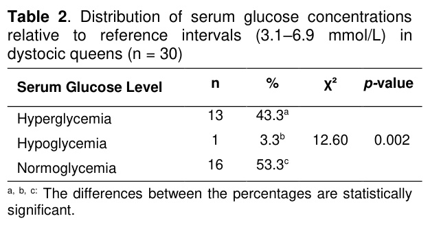
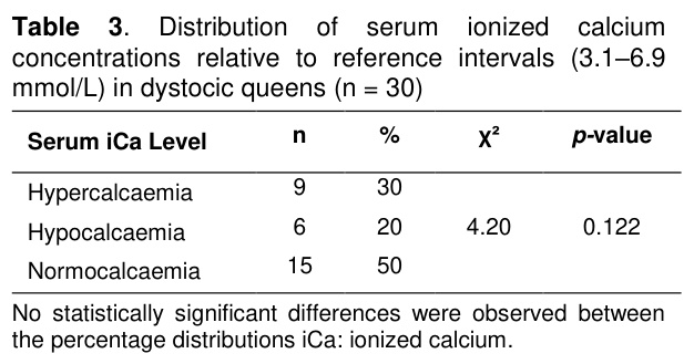
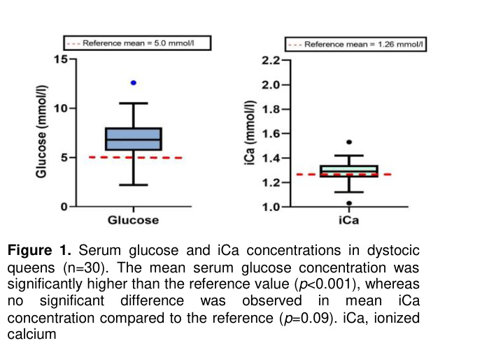
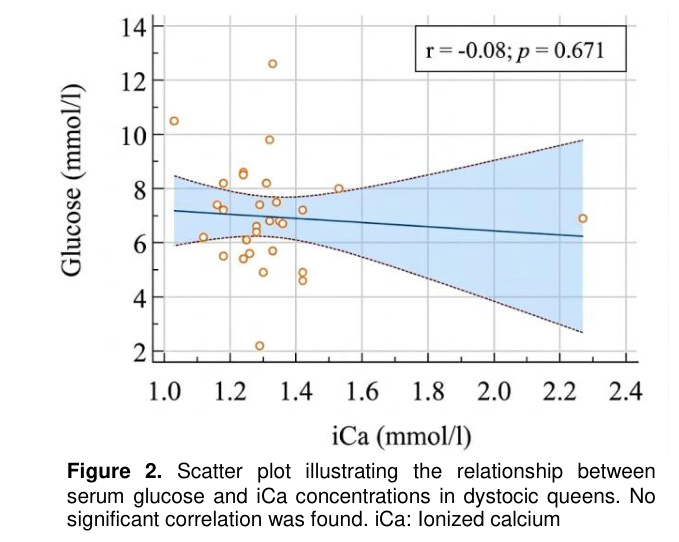
Descriptive statistics for serum glucose and iCa concentrations in dystocic queens are presented in Table 1. The mean serum glucose and iCa values were 6.96±1.94 mmol/L and 1.31±0.20 mmol/L, respectively. Compared to the established reference mean for glucose (5.00 mmol/L [16]), serum glucose concentrations were significantly elevated in dystocic queens (p<0.001). In contrast, the mean iCa concentration did not differ significantly from the reference value of 1.26 mmol/L (p=0.09) (Figure 1). Based on the reference interval for blood glucose (3.1–6.9 mmol/L; [16]), 43.3% of queens (13/30) were classified as hyperglycemic, 53.3% (16/30) as normoglycemic, and 3.3% (1/30) as hypoglycemic. The distribution of glucose values significantly deviated from the expected reference distribution (p=0.002; Table 2). With respect to serum iCa reference values (1.20–1.32 mmol/L [16]), 30% (9/30) of queens exhibited hypercalcemia, 50% (15/30) normocalcemia, and 20% (6/30) hypocalcemia. However, these proportions did not show a statistically significant deviation (p=0.122; Table 3). Correlation analysis between serum glucose and iCa concentrations revealed a weak, non-significant negative relationship (r=–0.08, p=0.671 Figure 2). Overall, these findings suggest that deviations in serum glucose and iCa concentrations are not major contributors to the etiology of dystocia in queens with uterine inertia.

Discussion
Currently, studies investigating blood glucose and iCa concentrations in dystocic queens are limited. A literature search was conducted using the PubMed database with the keywords “blood glucose” and “iCa levels in dystocic queens in (2025). This search yielded only two relevant studies on the topic, each proposing distinct hypotheses [8,15]. No other publications specifically addressing this association were identified. Therefore, this study aimed to determine blood glucose levels and serum iCa concentrations in dystocic queens. The findings may help address the current gap in the literature regarding the role of glucose and calcium in the etiopathogenesis of dystocia in queens. Furthermore, the present study appears to be among the first to examine the correlation between blood glucose and iCa concentrations in dystocic queens.Among the maternal factors contributing to dystocia in queens, uterine inertia is recognized as one of the most prominent etiological causes [15,17]. This phenomenon observed in bitches may result from insufficient calcium levels, which are essential for uterine muscle contractility, and inadequate glucose availability, which compromises the energy demands of parturition [1,9,10,18]. Research conducted on both humans and rodent models underscores the pivotal role of calcium in modulating myometrial contractility during parturition. Importantly, calcium mobilization from the myometrial sarcoplasmic reticulum is characterized by its transient nature and rapid depletion, contrasting with the more sustained availability of extracellular calcium. Conversely, calcium influx serves as a critical driver of the rhythmic depolarization observed in pacemaker regions of uterine myocytes, thereby orchestrating synchronized myometrial contractions [19,20].
In many studies, the diagnosis of uterine inertia has been based on anamnestic data, behavioral observation of the queen, or the response to oxytocin administration [8,15,17]. However, external tocodynamometry is widely used in clinical practice due to its noninvasive nature and its ability to accurately time uterine contractions in relation to fetal heart rate monitoring [21]. In our study, other potential causes of dystocia such as transverse fetal positioning or fetal macrosomia were excluded through ultrasonographic and radiographic evaluation.
Normal parturition relies on the presence of regular and effective contractions of the uterine myometrium. The mechanisms responsible for initiating and sustaining the synchronized uterine activity necessary for delivery in queens involve a complex interplay of hormonal and mechanical factors that remain incompletely understood. Calcium is thought to play a pivotal role in uterine smooth muscle contraction, while glucose serves as a key metabolic substrate during labor [11]. A recent study [15], for the first time, challenged this conventional understanding by reporting no association between dystocia and hypocalcemia or hypoglycemia in queens. Conversely, another recent investigation [8] demonstrated that exogenous calcium supplementation may be beneficial in dystocic queens with hypocalcemia. In contrast to these previous reports, which were based on relatively small cohorts [n=19 [15] and n=8 for uterine inertia [8]], our study utilized a significantly larger group of 30 queens, providing a more robust statistical basis for evaluating these metabolic parameters. Notably, this conclusion was drawn from a limited subset of only 8 queens (8.9%) diagnosed with uterine inertia. Furthermore, in the same study, 4 dystocic queens with normocalcemic profiles also responded positively to calcium therapy, suggesting that calcium may contribute to uterine contractility independent of circulating iCa levels. Interestingly, it has been reported that hypercalcemia occurred in 57.9% of dystocic queens, with no cases of hypocalcemia observed [15]. In our study, which included a larger number of dystocic queens, the incidence of hypercalcemia was 30%, and hypocalcemia was identified in 20% of cases. In contrast, another study reported relatively low rates of hypocalcemia (8.9%) and hypercalcemia (6.7%) among the subjects evaluated [8]. These discrepancies in iCa distribution across studies may partly stem from differences in the reference intervals used to define normal iCa levels in cats. For instance, a previous study [22] reported a reference range of 1.15–1.37 mmol/L in 92 healthy cats and noted a significant age-related decline in blood iCa concentrations. This variability underscores the need for age-adjusted and standardized reference values when interpreting calcium status in feline obstetrics. Thus, in the absence of age-specific reference intervals, clinical interpretation of serum iCa deviations may be limited, especially in feline populations with heterogeneous age distributions. As a limitation of our study, since it did not include a control group of queens undergoing normal parturition (eutocia), blood glucose and iCa concentrations in dystocic queens were compared to reference values from healthy non-pregnant queens. However, it should be noted that factors such as age, breed, reproductive status, and stress may also influence blood reference values in healthy queens.
Increased calcium demands for fetal mineralization during late pregnancy may result in maternal hypocalcemia, which can contribute to dystocia due to reduced myometrial contractility [11]. However, contrary to this classical view, a previous study [15] reported that hypocalcemia is not a primary cause of dystocia in queens. Consistent with this, our study found no statistically significant differences between serum iCa concentrations and reference values. These findings contrast with earlier reports [8], suggesting that serum iCa concentrations are not a definitive marker in the etiology of dystocia associated with uterine inertia in queens. Nevertheless, hypocalcemia was identified in 20% of the queens included in the present study. Given the well-established role of calcium in uterine smooth muscle contraction, this proportion may still be clinically relevant in individual animals. Therefore, although our findings do not support hypocalcemia as a consistent etiological factor in dystocic queens with uterine inertia, they should not be interpreted as discouraging calcium supplementation in selected cases with documented or suspected calcium deficiency. Clinical decisions regarding calcium administration should be based on individual patient assessment and the overall obstetrical presentation. Consequently, the routine administration of calcium to all dystocic queens remains controversial, whereas targeted supplementation in appropriately selected cases may still be justified.
It has been reported that 65.5% of dystocic queens with uterine inertia exhibited hyperglycemia, with a positive correlation found between hyperglycemia and fetal mortality [8]. In our study, 43.3% of dystocic queens were hyperglycemic, whereas only one individual showed hypoglycemia. Serum glucose concentrations significantly differed from the reference mean (p=0.002). However, this finding should be interpreted with caution. Transient hyperglycemia has been previously associated with stress responses, such as cortisol release during parturition or veterinary examinations [23]. Similarly, a study in women demonstrated that elevated stress levels during labor can increase insulin requirements and result in higher blood glucose concentrations [24]. Similarly, another study [25] reported stress-induced elevations in blood glucose levels in dystocic bitches. Furthermore, Rand et al. [26] demonstrated that acute stress can induce marked but transient hyperglycemia in healthy cats, and that this response is associated with behavioral stress and increased norepinephrine concentrations. Similarly, a study in women demonstrated that elevated stress levels during labor can increase insulin requirements and result in higher blood glucose concentrations [24]. Likewise, Lúcio et al. [25] reported stress-associated elevations in blood glucose concentrations in dystocic bitches [25]. More recently, Song et al. [27] summarized the mechanisms underlying stress-induced hyperglycemia and emphasized the roles of catecholamines, cortisol, inflammatory mediators, increased hepatic glucose production, and insulin resistance during acute stress conditions. Therefore, although a direct causal relationship could not be established in the present study, stress-related hyperglycemia may represent a plausible explanation for the elevated glucose concentrations observed in some dystocic queens with uterine inertia.
Taken together, our findings suggest that neither blood glucose nor iCa concentrations are definitive markers for diagnosing or explaining the etiology of uterine inertia in dystocic queens. Furthermore, the lack of a significant correlation between blood glucose and iCa levels in dystocic queens observed in our study should be interpreted with caution. This is because confirming the clinical relevance of this nonsignificant correlation would require future studies involving a substantially larger number of dystocia cases. The contradictory results reported in the literature highlight the need for further prospective studies with standardized reference ranges, larger sample sizes, and age-specific evaluations to clarify the role of these parameters in feline dystocia.
Conclusion
In conclusion, the findings of this study indicate that serum glucose and iCa concentrations may vary in dystocic queens with uterine inertia, although such fluctuations are not consistent or predictive across cases. Hypoglycemia and hypocalcemia were observed only in a small proportion of animals. Moreover, no correlation was found between serum glucose and iCa levels, suggesting that these parameters are independent of each other. Overall, our results suggest that deviations in blood glucose and iCa concentrations do not play a primary role in the pathogenesis of dystocia associated with uterine inertia in queens. These findings support the notion that dystocia is a multifactorial condition, and that glucose and calcium levels alone are insufficient as diagnostic or prognostic markers in its management. However, it may be premature to generalize these findings to all dystocic queens at this stage. Numerous factors that may influence the blood parameters investigated such as breed, age, stage of pregnancy, parity, body weight, temperament, environmental conditions, and the time elapsed before presentation to the clinic could potentially lead to varying results. Therefore, further similar studies are warranted to elucidate these aspects more comprehensively. As a retrospective study, the present investigation is subject to the limitations inherent to retrospective data collection, including potential selection bias. Although only cases with complete clinical and laboratory records were included and strict inclusion criteria were applied, the possibility of residual bias cannot be entirely excluded. The present study did not evaluate the potential effects of litter size, fetal viability, or duration of labour on serum glucose and ionized calcium concentrations. These factors may influence the clinical course of dystocia and could potentially affect metabolic parameters. Therefore, future prospective studies incorporating these variables may provide a more comprehensive understanding of their role in dystocic queens with uterine inertia. Another limitation of the present study is the absence of a control group of queens undergoing normal parturition. Consequently, serum glucose and ionized calcium concentrations in dystocic queens were compared with published reference values rather than with contemporaneously evaluated eutocic queens. Future prospective studies including matched control populations would provide more robust evidence regarding the role of these parameters in feline dystocia. An additional limitation of this study is that primary and secondary uterine inertia could not be unequivocally differentiated in all cases because of the retrospective study design. Although all queens were presented during stage 2 labour and most cases were clinically consistent with secondary uterine inertia, the possibility that some cases represented primary uterine inertia cannot be completely excluded.References
- Jutkowitz LA. Reproductive emergencies. Vet Clin North Am Small Anim Pract 2005; 35: 397-420.
- Sendag S, Hardegen D, Koca D, Wehrend A. Investigation of maternal mortality and stillbirth in feline dystocia after emergency obstetric care interventions: A retrospective analysis. J Feline Med Surg 2025; 27: 1098612X241297898.
- Colby ED, Stein B. The Reproduction System: Feline Medicine. 1st Edition, American Veterinary Publications: California, 1983.
- Ekstrand C, Linde-Forsberg C. Dystocia in the cat: A retrospective study of 155 cases. J Small Anim Pract 1994; 3: 459-464.
- Gunn-Moore DA, Thrusfield MV. Feline dystocia: Prevalence, and association with cranial conformation and breed. Vet Rec 1995; 136(14): 350-353.
- Gendler A, Brourman JD, Graf KE. Konservative und operative Behandlung der Dystokie beim Hund. Tierarztl Prax Ausg K Kleintiere Heimtiere 2008; 36: 55-64.
- Sendag S, Wehrend A. Untersuchung zur Beziehung zwischen gestationsdauer und alter, körpergewicht bzw. wurfgröße bei van-katzen. Tierarztl Prax Ausg K Kleintiere Heimtiere 2010; 38: 163-168.
- Axnér E, Jakobsson J, Vermelin T, Hermansson U. A retrospective study on dystocia in the cat: Evaluation of 111 cases. Acta Vet Scand 2025; 67(1): 20.
- Bergström A, Fransson B, Lagerstedt AS, Olsson K. Primary uterine inertia in 27 bitches: Aetiology and treatment. J Small Anim Pract 2006; 47(8): 456-460.
- Hollinshead FK, Hanlon DW, Gilbert RO, et al. Calcium, parathyroid hormone, oxytocin and pH profiles in the whelping bitch. Theriogenology 2010; 73(9): 1276-1283.
- Reichler I, Michel E, Hubler M. Trächtigkeit und Geburt. In: Lutz H, Kohn B, Forterre F (Editors). Krankheiten der Katze. 6th Edition. Thieme: Stuttgart, 2019.
- Johnson SD. Management of Pregnancy Disorders in the Bitch and Queen. 1st Edition, WB Saunders Co: Philadelphia, 1983.
- Linde-Forsberg C, Eneroth A. Parturition. In: Simpson GM (Editor). BSAVA Manual of Small Animal Reproduction and Neonatology. 1st Edition, BSAVA: London, 1998.
- Linde-Forsberg C. Abnormalities in pregnancy, parturition and the periparturient period. In: Ettinger SJ, Feldman EC (Editors). Textbook of Veterinary Internal Medicine. 6th Edition, Elsevier Saunders: St. Louis, 2005.
- Bailin HG, Thomas L, Levy NA. Retrospective evaluation of feline dystocia: Clinicopathologic findings and neonatal outcomes in 35 cases (2009–2020). J Feline Med Surg 2022; 24(4): 344-350.
- Kraft W, Dürr UM. Klinische Labordiagnostik in der Tiermedizin. 5th Edition, Schattauer: New York, 1999.
- Holst BS, Axnér E, Öhlund M, Möller L, Egenvall A. Dystocia in the cat evaluated using an insurance database. J Feline Med Surg 2017; 19(1): 42-47.
- Frehner BL, Reichler IM, Keller S, Goericke-Pesch S, Balogh O. Blood calcium, glucose and haematology profiles of parturient bitches diagnosed with uterine inertia or obstructive dystocia. Reprod Domest Anim 2018; 53(3): 680-687.
- Testrow CP, Holden AV, Shmygol A, Zhang H. A computational model of excitation and contraction in uterine myocytes from the pregnant rat. Sci Rep 2018; 8(1): 9159.
- Malik M, Roh M, England SK. Uterine contractions in rodent models and humans. Acta Physiol 2021; 231(4): e13607.
- Rosen H, Yogev Y. Assessment of uterine contractions in labor and delivery. Am J Obstet Gynecol 2023; 228(5): 1209-1221.
- Deniz A, Mischke R. Ionisiertes calcium und Gesamtcalcium bei der Katze. Berl Munch Tierarztl Wochenschr 1995; 108: 105-108.
- Mischke R. Anämie. In: Mischke R (Editor). Praktische Hämatologie bei Hund und Katze. 1st Edition, Schlütersche Verlag: Hannover, 2003.
- Jovanovic L. Glucose and insulin requirements during labor and delivery: The case for normoglycemia in pregnancies complicated by diabetes. Endocr Pract 2004; 10: 40-45.
- Lúcio CF, Silva LCGD, Vannucchi CI. Perinatal cortisol and blood glucose concentrations in bitches and neonatal puppies: Effects of mode of whelping. Domest Anim Endocrinol 2021; 74: 106483.
- Rand J, Kinnaird E, Baglioni A, Blackshaw J, Priest J. Acute stress hyperglycemia in cats is associated with struggling and increased concentrations of lactate and norepinephrine. J Vet Intern Med 2002; 16(2): 123-132.
- Song G, Liu X, Lu Z, et al. Relationship between stress hyperglycaemic ratio (SHR) and critical illness: A systematic review. Cardiovasc Diabetol 2025; 24: 188.