
Kadmiyumun Neden Olduğu Karaciğer Dokusu Hasarında Linalool'un Spexin Düzeyleri ve İnflamasyon Üzerine Etkisi
Volume 38, Issue 1
January 2024 · Pages 062-069
Kadmiyumun Neden Olduğu Karaciğer Dokusu Hasarında Linalool'un Spexin Düzeyleri ve İnflamasyon Üzerine Etkisi
Effect of Linalool on Spexin Levels and Inflammation in Cadmium-Induced Liver Tissue Damage
Sercan KAYA1, Tuba YALÇIN1
Amaç: Çevre kirletici bir ağır metal olan kadmiyum (Cd), başta böbrek ve karaciğer olmak üzere birçok dokuda birikerek toksik etkiler gösterir. Doğal ürünlerin uçucu yağlarında bulunan ve parfümeri/kozmetik alanlarında sıklıkla kullanılan linalool’ün (LIN), ilginç bir şekilde anti-mikrobiyal, anti-kanser, anti-inflamatuvar gibi yararlı özellikler sergilediği yakın zamanda belirlenmiştir. Bu çalışma, Cd kaynaklı karaciğer dokusu hasarına karşı LIN uygulamasının, nöropeptit Speksin (SpX) ve inflamasyonu düzenleyici potansiyel etkisini belirlemeyi amaçladı.
Gereç ve Yöntem: Çalışmada 28 adet sıçan rastgele eşit sayıda 4 gruba ayrıldı. Gruplar; kontrol (n=7), Cd ( n=7; ilk 7 gün 3 mg/kg CdCl2), Cd+LlN (n=7; ilk 7 gün 3 mg/kg Cd ve 100 mg/kg/gün LIN) ve LIN (n=7; 100 mg/kg/gün LIN) olacak şekilde tasarlandı. 15. gün deney sonlandırıldı. Alınan kan serumu örnekleri biyokimyasal analizlerde ve karaciğer dokuları histopatolojik ve immünohistokimyasal analizlerde kullanıldı.
Bulgular: Cd kaynaklı karaciğer enzim düzeyleri, apoptotik ve inflamatuvar belirteçlerin immünreaktiviteleri artarken SpX immünreaktivitesi karaciğerde azalmıştı. Ayrıca, LIN uygulamasının Cd maruziyetine bağlı ortaya çıkan olumsuzlukları düzenleyici etkisi olduğu tespit edildi.
Sonuç: Cd kaynaklı karaciğer dokusu hasarına karşı LIN, SpX ve inflamasyonu düzenleyerek hepatoprotektif bir etki gösterebilir.
Aim: Cadmium (Cd), an environmentally polluting heavy metal, accumulates in many tissues, especially the kidney and liver, and has toxic effects. It has recently been determined that linalool (LIN), which is found in the essential oils of natural products and frequently used in perfumery/cosmetics, interestingly exhibits beneficial properties such as anti-microbial, anti-cancer and anti-inflammatory effects. This study aimed to determine the potential effect of LIN application on regulating neuropeptide Spexin (SpX) and inflammation against Cd-induced liver tissue damage.
Materials and Methods: In the study, 28 rats were randomly divided into 4 equal groups. The groups were; control (n=7), Cd (n=7; 3 mg/kg CdCl2 for the first 7 days), Cd+LlN (n=7; 3 mg/kg CdCl2 for the first 7 days and 100 mg/kg/day LIN) and LIN (n=7; 100 mg/kg/day LIN). The experiment was terminated on the 15th day. The blood serum samples taken were used in biochemical analyzes and the liver tissues were used in histopathological and immunohistochemical analyses.
Results: While liver enzyme levels and immunoreactivities of apoptotic and inflammatory markers increased due to Cd exposure, SpX immunoreactivity decreased in the liver. Additionally, it was detected that LIN application had a regulatory effect on the negative effects caused by Cd exposure.
Conclusion: Against Cd-induced liver tissue damage, LIN may exert a hepatoprotective effect by regulating SpX and inflammation.
Introduction
Cadmium (Cd), one of the heavy metals, is an environmental pollutant with toxic effects[1]. The most obvious toxic property of CD is its extraordinarily long half-life in the human body. After Cd is absorbed, it accumulates at high levels in the human body [2]. Cd accumulates mainly in tissues such as kidney and liver and binds to the apoprotein metallothionein (MT). When the amount of Cd exposure surpasses intercellular MT levels, free Cd ions (Cd+2) that are not bound to MT in the cells cause inflammation, free radical formation, cell apoptosis, ultimately causes toxicity[3]. Although hepatic cells and proximal tubules of nephrons are the two main sites of Cd toxicity, hepatocellular damage is much greater than renal damage in the early stages of Cd exposure[4]. Cd is transported to the liver tissue by binding to the cellular membrane and via membrane transporters found in sinusoidal endothelial cells[5]. Therefore, inflammation and apoptosis are two important biomarkers in the liver damage process of Cd exposure[6].Natural products such as essential oils and their components are widely used due to their various bioactivities. Linalool (LIN) is an aromatic monoterpene alcohol found in essential oils and widely used in fields such as perfumery, cosmetics, food additives[7]. LIN is mainly obtained through chemical synthesis[8]. In recent years, it has been known that LIN has been injected into the inner space (food-contact space) of food packages[9]. In addition, LIN is used in the pharmaceutical industry as a precursor of vitamin E synthesis[10]. Interestingly, it has recently been reported that LIN exhibits bioactive properties such as anticancer, antimicrobial, neuroprotective, and hepatoprotective effects. It has also been reported that the preventive effect of LIN in kidney and liver tissues is due to its anti-inflammatory activity[7].
Spexin (SpX; Neuropeptide Q), a recently identified peptide, is expressed in many tissues, such as liver, kidney, lung, and central nervous system[11]. SpX is an associate of the galanin (GAL)/kisspeptin family of peptides. It shows its biological activities through GAL receptors (GALR). Interestingly, GAL exhibits greater affinity for the receptors GALR1 and GALR2, whereas SpX exhibits greater affinity for GALR2 and GALR3[12]. Endogenous neuropeptide SPX plays an important role in the regulation and control of many physiological functions such as energy metabolism, gastrointestinal system, kidney and cardiovascular functions[13]. However, many studies have shown that SpX is associated with pathological conditions such as type 2 diabetes and obesity[14-16]. Additionally, SpX has been reported to have a role in inflammatory processes, but this needs to be further clarified[17].
To our knowledge, there has been no previous study on the potential therapeutic effect of LIN against liver tissue damage due caused by Cd exposure and how SpX levels are affected in this process. Therefore, this study was conducted to appraise the anti-inflammatory and SpX levels regulatory potential of LIN application against Cd-induced hepatotoxicity.
Material and Method
Research and Publication Ethics: This study was approved by the decision of Fırat University Animal Experiments Ethics Committee dated 22.09.2023-18332.Experimental Design: The 28 Sprague-Dawley male rats (180-210 g/8-10 weeks) used in this study were housed under appropriate conditions (12 h light/dark cycle, 22-24°C, water and food adlibitum). The rats used in the study were randomly divided into 4 groups, with equal numbers of rats in each group (n=7). Control group; no treatment was applied to the rats in this group. Cd group; Cd (CdCl2-20290810108-64-2, Sigma-Aldrich) was given at a dose of 3 mg/kg for the first 7 days of the experiment. Cd+LlN group; For the first 7 days of the experiment Cd at a dose of 3 mg/kg and LIN at a dose of 100 mg/kg/day (97%, L2602-102524372, Sigma-Aldrich) were administered. LIN group; LIN was administered at a dose of 100 mg/kg/day. No deaths or adverse events were reported during the experiment. Cd[18] and LIN[19] doses were taken as reference from previous studies. Cd and LIN were dissolved in saline solution and all treatments were administered intraperitoneally. At the end of the experiment (15th day), all rats were decapitated under anesthesia (ketamine/xylazine-75/10 mg/kg). Blood serums and liver tissues were collected quickly and stored at -80°C until the day of study for biochemical, histopathological and immunohistochemical examinations.
Determination of Alanine aminotransferase and Aspartate Aminotransferase levels: Liver enzyme levels in serum samples were analysed by using ADVIA 2400 autoanalyser device (Siemens, Germany) and kits in the central laboratory of Fırat University.
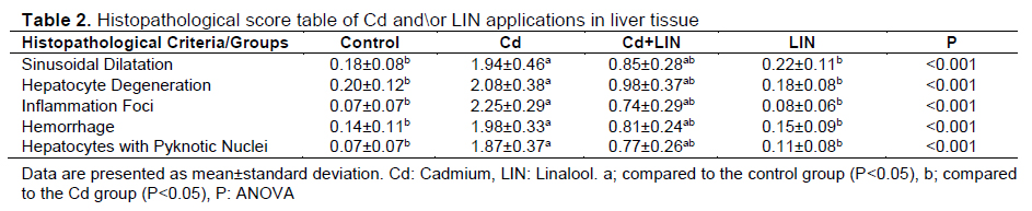
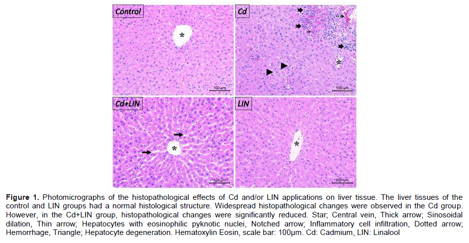
Histopathological Evaluations: After collected, the liver tissues were placed in 10% formalin and subjected to routine histological follow-up series after fixation. Hematoxylin-Eosin staining procedure was applied to liver tissue sections for histopathological evaluation. The liver tissue preparations were assessed with a light microscope and microphotographs were taken (DM2500/Leica, Germany). Evaluation criteria; sinosoidal dilatation, hepatocyte degeneration, inflammatory cell infiltration, hepatocytes with eosiniphilic pyknotic nuclei and the presence of hemorrhagic areas. Histopathological score (Table 2) was generated considering the presence/absence of histopathological criteria (0; none, 1; mild, 2; moderate and 3; excessive)[20].
Immunohistochemical Examinations: In liver tissue sections, Interleukin 1β (IL-1β) (1:200, Santa Cruz, sc-1251, USA), Cysteine-aspartic protease 3 (Casp-3) (1:200, Bioss, bs0081R, China), SpX (1:200, Boster, A04088-1, USA) and Tumor Necrosis Factor Alpha (TNF-α) (1:100, Elabscience, BL3376, USA) immunoreactivities were detected by the Avidin Biotin Peroxidase Complex method as previously reported[21]. The sections taken from the paraffin blocks with a thickness of 4–5 μm were placed on slides and deparaffinized. Then, the sections were passed through alcohol series and boiled in citrate buffer solution at pH:6 in a microwave oven (750 W) for 12 minutes. After boiling, the tissues were kept at room temperature to cool and washed with PBS (Phosphate Buffered Saline) and then hydrogen peroxide solution was applied for 6 minutes to prevent endogenous peroxidase activity. After applying block solution for 5 minutes to the tissues, which were washed with PBS for 3x5 minutes, they were incubated with primary antibodies 60 minutes in a humid environment at room temperature. After the application of the primary antibody, the tissues were washed with PBS for 3x5 minutes and then incubated with the secondary antibody compatible with the primary antibody for 30 minutes in a humid environment at room temperature. The chromogen utilized was 3-amino-9-ethyl carbazole (AEC) (TA-060-HA, Thermo Sci., England). Mayer Hematoxylin was used to counterstain all sections. The prepared preparations were evaluated with a light microscope and microphotographs were taken (DM2500Leica, Germany). Immunoreactivity: prevalence (<25%= 0.1, 26-50%= 0.4, 51-75%= 0.6, 76-100%= 0.9) X severity (none= 0; very little= 0.5; little= 1; moderate= 2; more= 3), It was calculated using the formula[22].
Statistical Analyzes: Statistical analyzes of the study data were performed using the SPSS 22.0 software program. Shapiro-Wilk test was used as a homogeneity test in statistical analyses. Parameters complying with normal distribution were analyzed with Oneway ANOVA post-hoc TUKEY tests. In the statistical analysis of parameters that did not comply with normal distribution, Kruskal Wallis followed by Mann Whitney U test was used. A value of P< 0.05 was considered statistical significance. Graphs of the study results were drawn using Graph Pad Prism 9.3.1.
Results
Effect of Cd and/or LIN Applications on Liver Enzyme Levels: Liver enzyme levels, which are frequently used to detect liver damage, were at similar levels in the control and LIN groups (P>0.05). It was determined that Alanin aminotransferaz (ALT) ve Aspartat Aminotransferaz (AST) levels increased in the Cd group compared to the control group (P<0.05). In the Cd+LIN group, ALT and AST levels decreased compared to the Cd group (P<0.05) (Table 1).

Effects of Cd and/or LIN Applications on Liver Histopathology: Liver tissues of the control and LIN groups exhibited normal histological structure. The presence of sinosoidal dilatation, hepatocyte degeneration, hemorrhagic areas, inflammation foci and hepatocytes with eosinophilic pyknotic nuclei were detected in the Cd group compared to the control group. It was observed that Cd-induced histopathological changes were significantly reduced in the Cd+LIN group (P<0.05) (Table 2, Figure
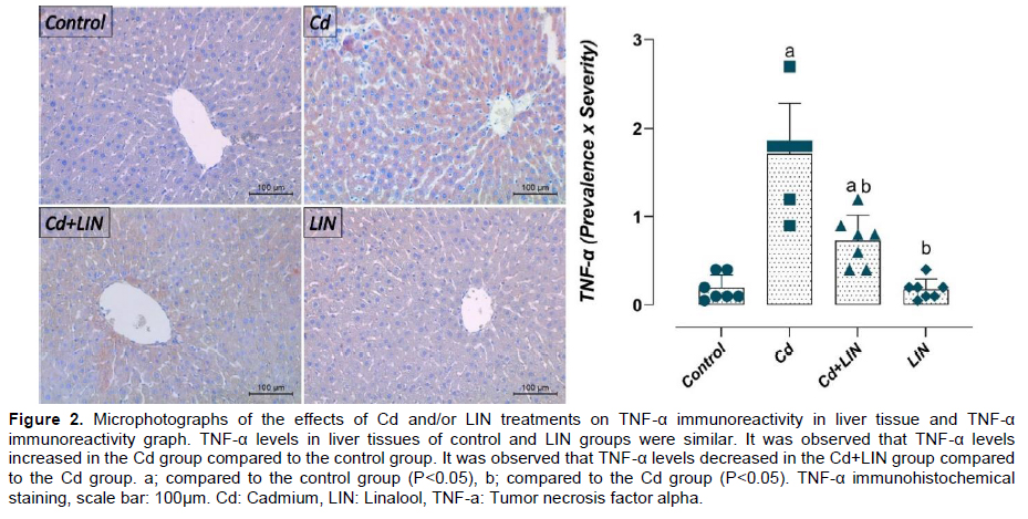
Effect of Cd and/or LIN Applications on IL-1β and TNF-α Immunoreactivities in liver: TNF-α immunoreactivity in the liver tissues of the control and LIN groups were similar (P>0.05). TNF-α immunoreactivity was increased in the Cd group compared to the control group (P<0.05). TNF-α immunoreactivity was decreased in the Cd+LIN group compared to the Cd group (P<0.05) (Figure 2).
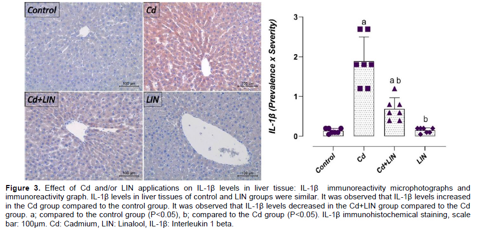
Similarly, IL-1β immunoreactivity was similar in liver tissues of control and LIN groups (P>0.05). IL-1β immunoreactivity was increased in the Cd group compared to the control group (P<0.05). However, it was determined that IL-1β immunoreactivity was decreased in the Cd+LIN group compared to the Cd group (P<0.05) (Figure 3).
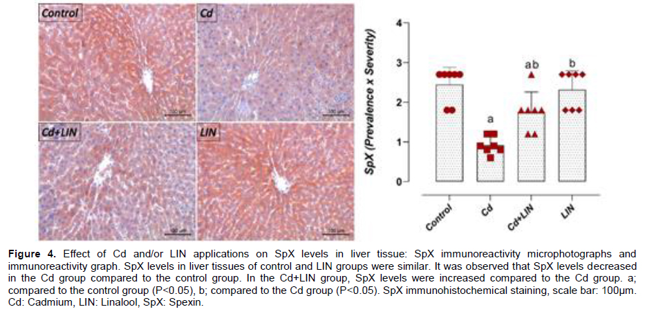
Effect of Cd and/or LIN Applications on SpX Immunoreactivities in Liver: SpX immunoreactivity in liver tissues of control and LIN groups was similar (P>0.05). SpX immunoreactivity was found to be increased in liver tissues in the Cd group compared to the control group (P<0.05). The Cd+LIN group showed decreased SpX immunoreactivity compared to the Cd group (P<0.05) (Figure 4).
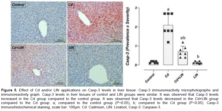
Effect of Cd and/or LIN Applications on Casp-3 Immunoreactivity in Liver: Liver tissues showed similar Casp-3 immunoreactivity in control and LIN groups (P>0.05). It was detected that Casp-3 immunoreactivity in liver tissues increased in the Cd group compared to the control group (P<0.05). A decrease in Casp-3 immunoreactivity was detected in the Cd+LIN group compared to the Cd group (P<0.05) (Figure 5).
The increase in AST and ALT levels, which are frequently used clinically as liver function tests, indicates the deterioration of membrane integrity in hepatocytes[26]. Therefore, AST and ALT are also used as markers of liver tissue damage. It has been shown in many studies that Cd exposure significantly increases liver enzymes[1,27,28]. In this current study, consistent with previous studies, it was found that Cd augmented liver enzyme levels. However, many studies have reported that Cd causes widespread histopathological changes in liver tissue[29,30]. For example, it has been reported that Cd exposure causes hepatocyte degeneration, inflammatory cell infiltration, dilated sinosoids and necrotic cells in liver tissue[1]. Similarly, in this current study, it was shown that Cd application caused histopathological changes in liver tissues. On the other hand, it was determined that LIN application had a regulatory effect on Cd-induced increased liver enzyme levels and histopathological changes. These results are coherent with previous studies reporting that LIN application exhibits regulatory effects on liver enzyme levels and histopathological changes in many different hepatotoxic models such as carbon tetrachloride[31], benzene [32], ischemia-reperfusion[33] and high-fat diet[34].
Cd accumulates in the liver and causes inflammatory reactions [28]. Many studies have confirmed that inflammation is associated with Cd-induced toxicity[35,36]. In response to Cd toxicity, resident macrophages and Kupffer cells in the liver are activated. This triggers a complex network of inflammatory mediators. Thus, proinflammatory cytokines are (IL-1β, TNF-α, and IL-6) secreted in the liver[37,38]ğ. In a study conducted on rats, Cd caused a significant decrease in the level of IL-10 (anti-inflammatory cytokine) while increasing pro-inflammatory cytokines (IL-1β, TNF-α and IL-6) in liver tissue[39]. Similarly, in this current study, IL-1β and TNF-α immunoreactivities were found to increase in Cd-induced liver tissue. The results obtained were consistent with previous studies reporting Cd-induced increases in pro-inflammatory cytokines in the liver[1,28,40]. One study reported that oleic acid has a liver-protective effect by reducing the levels of pro-inflammatory cytokines, which increase due to Cd[40]. LIN was reported to have a protective effect against peripheral inflammation caused by the endotoxin Salmonella typhimurium[41]. In addition, LIN suppressed nephropathy by exhibiting anti-inflammatory and antioxidant effects in a diabetic rat model[42]. However, LIN also inhibited TNF-α-induced inflammation in brain tissue endothelial cells[43]. It has also been reported that LIN inhibits proinflammatory pathways and cytokines induced by LPS[44,45]. Similarly, in this current study, LIN reduced the immunoreactivities of pro-inflammatory cytokines (TNF-α and IL-1β), which increased in Cd-induced liver tissue.
In homeostasis, adipokines (Adiponectin, Transforming growth factor beta, and IL10,) secreted by immune cells and adipocytes maintain physiological function, increase insulin sensitivity and inhibit inflammation in adipose tissues. The inflammatory environment that develops in fat tissues with obesity increases with the secretion of adipokines (IL6, resistin, leptin) released by adipocytes, infiltrated macrophages (M1) that differentiate into type 1 phenotype, and proinflammatory cytokines (IL6, TNFα and IL1β) by other inflammatory cells[17,46,47]. However, immune system cells (monocytes, neutrophils and macrophages) are known to express GALRs and play a role in inflammation[48]. It has been shown that SpX modulates inflammation in adipose tissues at the mRNA level when fed a fructose-rich diet and can improve the M1/M2 ratio by reducing the percentage of M1 macrophages[49]. In this study, SpX immunoreactivity decreased in Cd-induced liver tissue. Similarly, a recent study reported that endogenous SpX is susceptible to metabolic changes and SpX levels decrease in diseases such as obesity and diabetes. Additionally, the same study reported that SpX treatment showed many positive effects on metabolism, including reduction of lipid accumulation, fat mass, and inflammation[50]. Another study reported that SpX administration reduced IL-6 and TNF-α levels as well as lipid content in the liver of an experimental obesity and diabetes model[51]. Additionally, SpX application has been shown to regulate negative effects such as hepatocyte inflammation, necrosis, or bleeding in rats[14]. The current study results showed that SpX levels decreased in contrast to increased liver enzyme levels, histopathological changes, and proinflammatory cytokines in liver tissue due to Cd exposure. However, while LIN application had a regulating effect on liver enzyme levels, histopathological changes and pro-inflammatory cytokine levels, it also increased the Cd-induced decreased SpX immunoreactivity in liver tissue.
Casp-3 is an important cytoplasmic pro-enzyme that causes irreversible apoptosis when activated[2]. Studies have shown that Cd significantly increases Casp-3 levels in liver tissue[1,27]. However, a recent study reviewed the protective potential of LIN in a rat model of isoproterenol induced myocardial infarction. According to the results of this study, LIN showed a cardioprotective effect by increasing the level of Bcl2 (antiapoptotic), decreasing the levels of Nuclear Factor kappa B cytokines, IL-1β, TNF-α and IL-6, and apoptotic markers (Casp-3, Casp-9 and Bax)[52]. Similarly, in this study, it was detected that the increased Casp-3 immunoreactivity in Cd-induced liver tissues decreased with LIN application.
In this study, it was determined that Cd treatment increased liver enzyme levels, histopathological changes, and levels of inflammatory and apoptotic markers in liver tissues. It was also observed that Cd application reduced SpX immunoreactivity in liver tissue. On the other hand, it was determined that all Cd-induced negativities were regulated by LIN. In Cd-induced hepatotoxicity, LIN application showed a hepatoprotective effect by regulating proinflammatory cytokines and SpX levels. These results indicate that LIN, a monoterpene, may be a potential therapeutic candidate against heavy metal toxicity, but further and comprehensive studies are needed to determine its detailed mechanisms.
Discussion
Cd exposure remains a global public health problem[23]. There is significant evidence that Cd exposure is associated with inflammation (neutrophil infiltration and Kupffer cell activation), non-alcoholic fatty liver disease, non-alcoholic steatohepatitis, and necrotic hepatocellular death in humans[24,25]. LIN found in natural essential oils shows many bioactive properties such as hepatoprotective, nephroprotective, anti-inflammatory, anticancer and antimicrobial. It has also been reported that LIN can be used as an adjuvant of antibiotics or anticancer drugs due to its protective effects[7]. This study provides important evidence that LIN application against Cd-induced liver tissue damage has a hepatoprotective effect by suppressing inflammation and regulating SpX immunoreactivity.References
- Pathological Observation of Experimentally-Induced Cadmium Toxication in Foetal and Neonatal Mouse Pups
- Effect of Chelating Agents Against to Kidney and Liver Damage to Causes of Cadmium
- Determination of Levels Lead and Cadmium at Wildlife Pigeon in Elazıg Region
- Pathological Examination of Experimentally-Induced Cadmium Toxication in Rabbits
- Invastigation of the Effects of Some Steroids on Acute and Chronic Cadmium Toxicity in Mice