
Koyunlarda Sistemik Doramektin Uygulamasının Aköz Gözyaşı Üretim Miktarı Üzerine Etkileri
Volume 40, Issue 3
October 2026 · Pages 227-232
Koyunlarda Sistemik Doramektin Uygulamasının Aköz Gözyaşı Üretim Miktarı Üzerine Etkileri
Effects of Systemic Doramectin Administration on Aqueous Tear Production Quantity in Sheep
Yakup KOCAMAN1, Latif Emrah YANMAZ2, İrem SUZAK KOCAMAN3, Ferda TURGUT4, Vildan ASLAN CANATAN4
2Mehmet Akif Ersoy University, Faculty of Veterinary Medicine, Department of Surgery, Burdur, TÜRKİYE
3Erciyes University, Faculty of Veterinary Medicine, Department of Internal Medicine, Kayseri, TÜRKİYE
4Çukurova University, Faculty of Veterinary Medicine, Department of Surgery, Adana, TÜRKİYE
Makrosiklik laktonlar koyunlarda yaygın olarak kullanılan antiparaziter ajanlardır. Avermektin sınıfı bir endektosid olan doramektin yaygın olarak kullanılmaktadır; ancak gözyaşı salgısı ve göz yüzeyi üzerindeki etkileri yeterince araştırılmamıştır. Bu nedenle, bu çalışma klinik olarak sağlıklı koyunlarda sistemik olarak uygulanan doramektinin gözyaşı üretimi ve seçilmiş göz yüzeyi parametreleri üzerindeki etkilerini değerlendirmeyi amaçlamıştır. Toplam 64 adet 2-4 yaş arası klinik olarak sağlıklı dişi Kıvırcık koyun çalışmaya dahil edilmiştir. Doramektin, 200 µg/kg dozunda deri altına uygulanmıştır. Gözyaşı üretimi, tedavi öncesinde ve uygulamadan 48 saat sonra Schirmer Gözyaşı Testi I (STT-I) kullanılarak değerlendirilmiş ve kuru göz şiddeti STT-I değerlerine göre sınıflandırılmıştır. Tedavi öncesi ve sonrası ölçümler Wilcoxon işaretli sıra testi kullanılarak karşılaştırıldı ve gözyaşı üretimi değişiklikleri ile kuru göz şiddeti arasındaki ilişki Spearman'ın sıra korelasyonu kullanılarak değerlendirildi. Ortalama STT-I değerleri, tedavi öncesi 16.50±5.82 mm/dk'dan tedavi sonrası 13.69±4.47 mm/dk'ya önemli ölçüde azaldı (p<0.001). Doramektin uygulamasından sonra kuru göz şiddeti önemli ölçüde artmıştır (p=0.001). STT-I değerlerindeki değişiklikler ile kuru göz şiddetindeki değişiklikler arasında orta derecede, negatif ve istatistiksel olarak anlamlı bir korelasyon gözlemlenmiştir (r=−0.458; p<0.001). Floresein boyama, hiçbir hayvanda kornea hasarı olmadığını ortaya koydu. Sonuç olarak, bu çalışma sistemik doramektin uygulamasının koyunlarda gözyaşı üretimini önemli ölçüde azalttığını göstermektedir. Bu bulgular, doramektinin aköz gözyaşı üretiminde kısa dönemli subklinik değişikliklerle ilişkili olabileceğini düşündürmekte ve klinik kullanım sonrasında gözyaşı sekresyonunun izlenmesinin önemini vurgulamaktadır.
Macrocyclic lactones are commonly used antiparasitic agents in sheep. Doramectin, an avermectin-class endectocide, is widely administered; however, its effects on tear secretion and the ocular surface have not yet been adequately studied. Therefore, this study aimed to evaluate the effects of systemically administered doramectin on tear production and selected ocular surface parameters in clinically healthy sheep. A total of 64 clinically healthy female Kıvırcık sheep aged 2–4 years were included. Doramectin was administered subcutaneously at a dose of 200 µg/kg. Tear production was assessed before treatment and 48 hours after administration using the Schirmer Tear Test I (STT-I), and dry eye severity was classified according to STT-I values. Pre- and post-treatment measurements were compared using the Wilcoxon signed-rank test, and the association between changes in tear production and dry eye severity was evaluated using Spearman’s rank correlation. Mean STT-I values decreased significantly from 16.50±5.82 mm/min before treatment to 13.69±4.47 mm/min after treatment (p<0.001). Dry eye severity increased significantly following doramectin administration (p=0.001). A moderate, negative, and statistically significant correlation was observed between changes in STT-I values and changes in dry eye severity (r=−0.458; p<0.001). Fluorescein staining revealed no corneal damage in any animal. In conclusion, this study demonstrates that systemic doramectin administration significantly reduces tear production in sheep. These findings suggest that doramectin may be associated with short-term subclinical changes in aqueous tear production, highlighting the importance of monitoring tear secretion following clinical use.
Introduction
Antiparasitic drugs belonging to the macrocyclic lactones (MLs) family comprise the avermectins (ivermectin, doramectin, abamectin, and eprinomectin) and the milbemycins (moxidectin), which share closely related mechanisms of action. These agents exert their antiparasitic effects primarily by binding to glutamate-gated chloride channels, inducing neurotoxicity in parasites through hyperpolarization of target neuromuscular cells. In addition to their primary parasitic targets, macrocyclic lactones are also capable of potentiating other chloride channels, including gamma-aminobutyric acid (GABA)–gated channels within the mammalian central nervous system. Owing to their high efficacy and broad spectrum of activity, macrocyclic lactones are widely used worldwide for the control of endo- and ectoparasites across multiple animal species [1]. MLs are generally well tolerated at therapeutic doses. However, neurological side effects have occasionally been reported in dogs carrying ATP-Binding Cassette Subfamily B Member 1 (ABCB1) gene mutations that reduce drug efflux from the central nervous system [2]. At present, there have been no documented cases of ABCB1- related toxicity in sheep.Doramectin is a broad-spectrum macrocyclic lactone endectocide belonging to the avermectin class, and it is widely used in sheep for the treatment and control of gastrointestinal nematode and lungworm infestations [3]. Although the primary mechanism of action of the MLs involves binding to glutamate- and γ-aminobutyric acid (GABA)–gated chloride channels in parasites, it has been reported that similar ligand-gated chloride channels are also expressed in mammalian tissues outside the nervous system, including various exocrine glands [4]. These receptors contribute to secretomotor control processes, and it has been suggested that their pharmacological modulation may theoretically alter glandular secretory activity. The lacrimal gland, which is responsible for maintaining the aqueous component of the tear film, exhibits marked sensitivity to autonomic and neurohumoral influences [5]. Even a transient suppression of parasympathetic activity may reduce tear flow, leading to disruption of ocular surface stability and the development of early-stage dry eye [6]. At present, there is no evidence to suggest that doramectin directly interacts with receptors in the lacrimal gland. In light of this, the observed changes in tear production are more likely to be mediated through neurophysiological mechanisms rather than resulting from a direct effect or cytotoxic damage to lacrimal tissue.
Although it is well established that certain systemic medications, such as anticholinergics, antihistamines, and sedatives, can reduce aqueous tear secretion [7], the ocular effects of antiparasitic agents have been only limitedly investigated from a scientific perspective. Among antiparasitic agents, macrocyclic lactones are of particular relevance, as a recent study in clinically healthy dogs demonstrated that systemic ivermectin administration reduced aqueous tear production and was accompanied by ocular surface-related changes [8]. Additionally MLs have been reported to induce various ocular adverse effects, including mydriasis, retinal dysfunction associated with photoreceptor damage, and even blindness [9-12]. These complications are rare and are observed predominantly following high-dose administrations [10,13]. Nevertheless, these findings indicate that macrocyclic lactones are capable of interacting with ocular tissues and may potentially influence secretory and/or neurophysiological pathways.
Considering doramectin’s high lipophilicity and extensive tissue distribution, together with the transient neurological effects observed during periods of peak plasma concentrations [10,14], it is biologically plausible that this drug may affect lacrimal gland function. In this context, the aim of the present study was to evaluate the effects of systemic doramectin administration at a standard therapeutic dose on tear production and ocular surface integrity in clinically healthy sheep. It was hypothesized that subcutaneous doramectin administration could lead to a measurable reduction in aqueous tear secretion, secondary to neurophysiological modulation of lacrimal gland function.
Material and Method
Research and Publication Ethics: This study was approved by the Local Ethics Committee of Yozgat Bozok University. (Decision No.: 1/2026, 04 Feb 2026).A total of 64 female Kıvırcık sheep, aged 2–4 years, were included in the study. Based on comprehensive general clinical examinations, together with ophthalmological, hematological, and biochemical evaluations, only clinically healthy animals with no evidence of ocular or systemic pathology were enrolled. In all sheep, the ocular adnexa were normal, corneal transparency was preserved, ocular reflexes were intact, and the animals tolerated manipulation during the Schirmer Tear Test I (STT-I) without the need for sedation.
Animals were excluded if they exhibited ocular abnormalities, a baseline STT-I value ≤5 mm/min, systemic disease, administration of any medication within the preceding seven days, prior anesthesia or sedation, a history of antiparasitic treatment other than doramectin, pregnancy, lactation, or unmanageable behavior.
Doramectin administration was performed not for experimental purposes, but as part of a routine antiparasitic prophylaxis program conducted under farm supervision. The study was therefore observational in design and aimed to evaluate ocular parameters following standard therapeutic treatment. No anesthesia, sedation, or additional invasive procedures were applied throughout the study period. All procedures were carried out in accordance with national regulations governing the protection of experimental animals.
Groups: Sheep in the post-doramectin (PostDRM) group received doramectin (Dectomax® 1%, Pfizer, USA) administered subcutaneously at a dose of 200 µg/kg within the preceding 48 hours, in accordance with the farm’s routine antiparasitic control program. The pre-doramectin (PreDRM) group consisted of sheep that had not received any antiparasitic treatment for at least 30 days. The doramectin dose was determined based on the therapeutic range accepted for the control of endo- and ectoparasites in sheep. The 48-hour interval was selected to correspond with the reported time frame during which doramectin reaches peak plasma concentrations [14].
Tear measurements were performed in each sheep at two time points: immediately before doramectin administration (baseline) and 48 hours after administration (post-treatment). The same time interval was applied in the PreDRM group to obtain comparable repeated measurements.
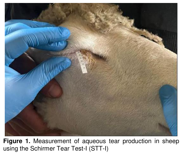
Tear Evaluation: Standard test strips were used for STT-I measurements (Schirmer test strip, No. 41 filter paper, 5 × 35 mm, ERC, Turkey). No topical anesthetics or sedatives were used. The strips were folded at the notched end and placed at the junction of the medial and middle thirds of the lower eyelid. After one minute, the length of the wetted area was recorded in millimeters per minute (mm/min) (Figure 1). In each sheep, only the right eye was included in the evaluation. This eye was selected in advance for all measurements to ensure consistency of the testing protocol and to avoid potential interference from reflex tearing that could be triggered in the opposite eye if STT-I were applied bilaterally. To minimize stress-induced lacrimation, all tests were performed in a quiet environment. All measurements were conducted under controlled environmental conditions, with a mean ambient temperature of 18.5 °C and relative humidity of 62%, to reduce potential variability in tear production. All ophthalmological assessments were conducted by the same investigator, who was blinded to the treatment status throughout the study. The severity of dry eye was graded according to STT-I values as follows: ≥15 mm/min was considered normal (grade 0), 11–14 mm/min mild (grade 1), 6–10 mm/min moderate (grade 2), and ≤5 mm/min severe (grade 3).
Assessment of Corneal Surface Health: Superficial corneal damage was evaluated using the fluorescein staining test. For each eye, 0.2 mL of a 1% fluorescein solution (Fluorescein, Kimyalab, Turkey) was instilled, and after one minute, excess dye was gently removed from the conjunctival sac using two drops of sterile 0.9% isotonic NaCl solution. The eyes were then examined under a focused light source.
Statistical Analysis: A sample size calculation was performed using G*Power software (version 3.1.9.7; Heinrich-Heine University Düsseldorf, Germany). Assuming a moderate effect size (Cohen’s dz = 0.5), an α level of 0.05, and 80% statistical power for a two-tailed paired-samples t-test, the minimum required sample size was calculated as 34 animals. In accordance with the pre–post study design, a total of 64 sheep were included, with measurements obtained from the same animals at both time points.
The distribution of continuous variables was assessed using the Kolmogorov–Smirnov test. As pre- and post-treatment measurements were obtained from the same animals, within-group comparisons were performed using either the paired-samples t-test or the Wilcoxon signed-rank test, depending on whether the differences followed a normal distribution. The relationship between changes in tear production and changes in dry eye severity was analyzed using Spearman’s rank correlation coefficient, based on the difference variables (ΔSTT and Δdry eye score).
All statistical analyses were conducted using IBM SPSS Statistics (version 26.0; IBM Corp., Armonk, NY, USA). All tests were two-tailed, and a p-value <0.05 was considered statistically significant. Data were presented as mean ± standard deviation (SD) or median (interquartile range, IQR) for continuous variables, and as median (IQR) for ordinal variables.
Results
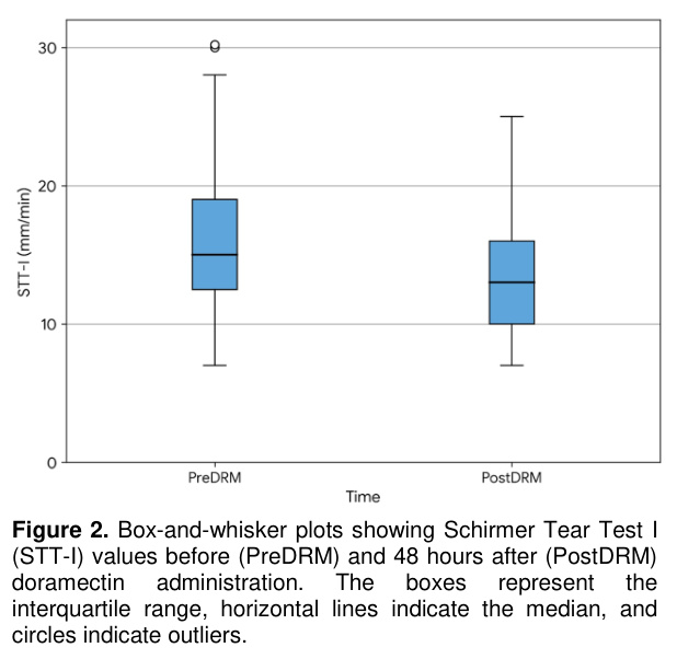
Before doramectin administration, the mean STT-I value in the sheep was 16.50±5.82 mm/min, whereas this value decreased to 13.69±4.47 mm/min after treatment (Table 1). As the distribution of the within-subject differences did not meet the assumption of normality, pre–post comparisons of STT-I values were performed using the nonparametric Wilcoxon signed-rank test. The Wilcoxon analysis demonstrated a statistically significant reduction in STT-I values following doramectin administration (Z = −5.584; p<0.001; Figures 2). STT-I values decreased in 50 animals, increased in 7 animals, and remained unchanged in 7 animals.Prior to doramectin administration, the median dry eye severity score was 0 (IQR: 1), whereas after treatment the median value increased to 1 (IQR: 2). As dry eye severity is an ordinal variable and did not follow a normal distribution, pre–post comparisons were conducted using the Wilcoxon signed-rank test. The analysis revealed a statistically significant increase in dry eye severity following doramectin administration (Z = −3.283; p=0.001). Dry eye severity increased in 20 animals, decreased in 4 animals, and remained unchanged in 40 animals (Table 1).
Spearman’s rank correlation analysis demonstrated a moderate, negative, and statistically significant association between ΔSTT and Δdry eye severity score (Spearman r= −0.458; p<0.001). This finding indicates that the post-doramectin reduction in tear production was associated with an increase in the severity of dry eye.
During fluorescein staining examinations, no corneal perforation was detected in any eye evaluated throughout the study period.
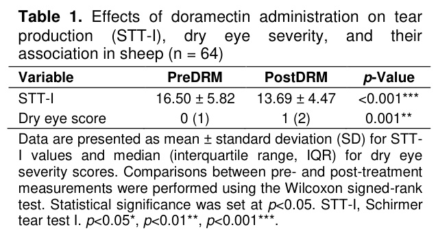
Data are presented as mean ± standard deviation (SD) for STT-I values and median (interquartile range, IQR) for dry eye severity scores. Comparisons between pre- and post-treatment measurements were performed using the Wilcoxon signed-rank test. Statistical significance was set at p<0.05. STT-I, Schirmer tear test I. p<0.05*, p<0.01**, p<0.001***.
Discussion
The primary objective of this study was to evaluate the effects of systemic doramectin administration at routine therapeutic doses on aqueous tear production in clinically healthy sheep. This study demonstrates that doramectin administration was associated with a significant short-term reduction in STT-I values in sheep. Following doramectin treatment, a statistically significant reduction in STT-I values was observed, accompanied by a significant increase in dry eye severity score. Moreover, the presence of a negative correlation between decreased tear production and increased dry eye severity indicates that these findings may be biologically related. In contrast, the absence of corneal damage on fluorescein staining in all cases suggests that doramectin did not induce detectable acute structural damage to the ocular surface within the 48-hour observation period. Taken together, these results indicate that doramectin administration may be associated with short-term subclinical changes in aqueous tear production in sheep and emphasize that tear secretion should be considered during the clinical use of long-acting macrocyclic lactones.STT-I values reflecting tear production in clinically healthy sheep have been reported in only a limited number of studies. In a study including different sheep breeds, Dedousi et al. reported a mean STT value of 18.45±3.93 mm/min and established a reference interval of 10.8–26.2 mm/min [15]. In line with these reference values, Gaffari et al. reported a mean STT-I value of 18.52±2.55 mm/min [16], Isler et al. reported 18.24±1.89 mm/min [17], and Akgül et al. [18] reported 18.83±5.29 mm/min for the right eye and 18.41±7.15 mm/min for the left eye. Accordingly, the STT-I values observed in the present study 16.50±5.82 mm/min in the PreDRM group and 13.69±4.47 mm/min in the PostDRM group remain within the physiological range.
Studies evaluating the effects of avermectins on tear production are limited. In a recent canine study, systemic ivermectin administration was associated with a significant reduction in aqueous tear production and a higher frequency of fluorescein uptake, supporting the possibility that avermectin-class macrocyclic lactones may influence tear-related ocular parameters [8]. By contrast, in a rabbit model, despite the development of marked ocular toxicity following systemic and intravitreal ivermectin administration, no statistically significant changes in Schirmer tear test values were observed [19]. Nevertheless, in the present study, systemically administered doramectin was found to significantly reduce aqueous tear production. The observed reduction in aqueous tear secretion 48 hours after doramectin administration may be attributable to the pharmacodynamic properties of the macrocyclic lactone used, the administered dose and route of administration, as well as species-specific physiological and anatomical differences.
Doramectin is defined as an antiparasitic agent that exerts its effects on γ-aminobutyric acid (GABA) receptors and glutamate-gated chloride ion channels involved in neuronal transmission in parasites, owing to its potential neurotoxic properties. In mammals, these ion channels are primarily localized within the central nervous system and are normally protected by the blood–brain barrier [20]. However, recent studies have demonstrated that GABAergic and glutamatergic receptors are also expressed in peripheral tissues outside the central nervous system, with notable expression in exocrine glands such as the lacrimal gland [21]. Activation of these inhibitory receptors may suppress parasympathetic neural transmission, thereby adversely affecting lacrimal gland secretion and leading to reduced tear production [22]. Within this framework, the decrease in STT-I values observed in the present study is considered to reflect a functional alteration in the neuroregulatory control of tear secretion, rather than indicating permanent or structural damage to the lacrimal gland parenchyma.
The plasma pharmacokinetic profiles of various endectocide molecules have been extensively characterized across different animal species in numerous studies [14]. It has been reported that the plasma elimination half-life of doramectin is approximately twice as long as that of ivermectin in cattle. Compared with ivermectin, doramectin has been shown to exhibit a more prolonged persistent activity in cattle, horses, pigs, and sheep, as demonstrated in multiple investigations [23]. In a study conducted by Imperiale et al. [14], doramectin administered subcutaneously to lactating sheep displayed long-term persistence in both plasma and milk, with measurable concentrations detected in both biological fluids for up to 30 days post-administration. Notably, the higher doramectin concentrations observed in milk compared with plasma indicate a marked tendency for accumulation within mammary gland tissue. In light of these findings, it should be considered that systemic doramectin administration may also result in prolonged retention within the lacrimal glands, potentially leading to subclinical dry eye manifestations and conferring a predisposition to the development of progressive keratoconjunctivitis sicca over time.
In sheep, doramectin is widely used for the treatment of psoroptic mange caused by Psoroptes ovis, an ectoparasitic disease that results in substantial economic losses in sheep farming worldwide [1]. Cantón et al. [1] reported the development of toxicity following iatrogenic doramectin overdose in sheep, with clinical manifestations including lethargy, aimless wandering, mydriasis, tachycardia, recumbency, and coma. The authors emphasized that animals in poor body condition and lambs are particularly predisposed to toxicity, and that at excessive doses, systemic drug concentrations may exceed the transport capacity of P-glycoprotein, thereby allowing macrocyclic lactones to cross the blood–brain barrier and enter the central nervous system. In contrast, in the present study, doramectin was administered within the reference therapeutic dose range for sheep, and no signs of toxicosis were observed. However, considering that doramectin may accumulate in adipose tissue in sheep with good body condition and maintain sustained plasma concentrations through slow release, it should be taken into account that the duration of subclinical dry eye may be prolonged.
Akgun et al. [24] reported that STT-I values decrease significantly as the severity of dry eye disease increases, and that this reduction in tear production is associated with ocular surface dysfunction. In parallel, the moderate, negative, and statistically significant correlation identified in the present study between ΔSTT and Δdry eye severity indicates that the reduction in tear volume after doramectin administration was accompanied by an increase in dry eye severity score. Accordingly, doramectin administration may influence aqueous tear production and may be associated with short-term changes in ocular surface-related parameters. This finding supports the importance of evaluating tear secretion during macrocyclic lactone use.
The present study has several limitations. First, the investigation focused solely on quantitative parameters reflecting tear volume, and other indicators of tear film quality, such as noninvasive tear break-up time, fluorescein tear break-up time, and tear osmolarity were not evaluated. Considering the lipophilic nature of doramectin and its potential persistence in milk and tissues for up to 30 days, future studies evaluating the long-term effects of macrocyclic lactones together with qualitative tear film tests such as TBUT would provide a more comprehensive understanding of their ocular effects. Therefore, the present findings should be interpreted as reflecting the acute phase only, and the persistence or reversibility of the observed reduction in tear production could not be determined within the scope of this study. Second, the assessment was limited to the short-term effects following systemic doramectin administration, and no histopathological or molecular analyses were performed to elucidate potential alterations in lacrimal gland function. In addition, the absence of fluorescein-positive corneal lesions at 48 hours does not exclude the possibility of delayed epithelial damage or subclinical dry eye related to impaired tear film stability. Third, the inclusion of animals from a single sheep breed restricted the exploration of breed-specific pharmacokinetic and physiological variations and their potential influence on tear-related parameters. Another limitation is the absence of a parallel untreated control group, which restricts the ability to fully exclude time-dependent physiological or environmental variations, although the within-subject design reduced inter-individual variability. In addition, comparative studies evaluating the ocular effects of doramectin and other macrocyclic lactones would be useful to better clarify whether these tear-related changes are specific to doramectin or represent a broader drug-class effect.
Conclusion
In conclusion, this study provides evidence that systemic doramectin administration in sheep is associated with a statistically significant short-term reduction in aqueous tear production. Although STT-I values decreased after treatment, most values remained within the physiological reference range; therefore, the clinical relevance of this reduction should be interpreted cautiously. The significant, moderate, and negative correlation between changes in STT-I values and dry eye severity score suggests a relationship between reduced tear production and increased dry eye grading after doramectin administration. In light of these findings, tear secretion may be monitored following systemic and long-acting doramectin administration, and the potential ocular effects of macrocyclic lactones should not be overlooked in clinical practice.References
- Cantón G, Urtizbiría F, Lloberas MM, et al. Iatrogenic doramectin overdosing causes toxicity in sheep: A case report. Vet Parasitol Reg Stud Reports 2022; 30: 100722.
- Mealey KL, Bentjen SA, Gay JM, Cantor GH. Ivermectin sensitivity in Collies is associated with a deletion mutation of the MDR1 gene. Pharmacogenet Genomics 2001; 11: 727-733.
- Gıcık Y, Arslan MÖ, Sarı B, Umur Ş. Efficacy of doramectin against gastrointestinal nematodes and lungworms in naturally infected pregnant sheep. Turkish Journal of Veterinary & Animal Sciences 2002; 26: 793-797.
- Merola VM, Eubig PA. Toxicology of avermectins and milbemycins (macrocylic lactones) and the role of P-glycoprotein in dogs and cats. Vet Clin North Am Small Anim Pract 2012; 42: 313-333.
- Dias-Teixeira K. Lacrimal gland postganglionic innervation: Unveiling the role of parasympathetic and sympathetic nerves in stimulating tear secretion. Am J Pathol 2020; 190: 968-969.
- Huang W, Tourmouzis K, Perry H, Honkanen R, Rigas B. Animal models of dry eye disease: Useful, varied and evolving (Review). Exp Ther Med 2021; 22: 1394.
- Kam KW, Di Zazzo A, De Gregorio C, et al. A review on drug-induced dry eye disease. Indian J Ophthalmol 2023; 71: 1263-1269.
- Kocaman Y, Yanmaz LE. Systemic ivermectin administration reduces aqueous tear production and affects ocular surface health in clinically healthy dogs. Vet Ophthalmol 2026; 29: e70180.
- Epstein SE, Hollingsworth SR. Ivermectin-induced blindness treated with intravenous lipid therapy in a dog. J Vet Emerg Crit Care 2013; 23: 58-62.
- Gwaltney-Brant SM, DeClementi C, Gupta RC. Macrocyclic lactone endectocides. In: Gupta RC (Editor). Veterinary Toxicology: Basic and Clinical Principles. 3rd Edition, London: Elsevier/Academic Press, 2018: 539-550.
- Kenny PJ, Vernau KM, Puschner B, Maggs DJ. Retinopathy associated with ivermectin toxicosis in two dogs. J Am Vet Med Assoc 2008; 233: 279-284.
- Merola VM, Eubig PA. Toxicology of avermectins and milbemycins (macrocyclic lactones) and the role of P-glycoprotein in dogs and cats. Vet Clin North Am Small Anim Pract 2018; 48: 991-1012.
- Simon AJ, Ferguson EGW. Dectomax injectable solution for cattle and sheep. Vet Rec 2004; 154: 341.
- Imperiale FA, Mottier L, Sallovitz JM, Lifschitz AL, Lanusse CE. Disposition of doramectin milk residues in lactating dairy sheep. J Agric Food Chem 2003; 51: 3185-3190.
- Dedousi A, Karatzia MA, Katsoulos PD. Reference values of Schirmer tear test in sheep and the effect of season on the test results. Acta Vet Hung 2019; 67: 553-560.
- Ghaffari MS, Shojaei M, Sabzevari A, Khorami N. Reference values for intraocular pressure and Schirmer tear test in clinically normal Sanjabi sheep. Small Rumin Res 2011; 97: 101-103.
- Işler CT, Altuğ ME, Kilic S. Evaluation of tear fluid secretion and intraocular pressure in normal Merinos sheep and Saanen goats. Rev Med Vet 2013; 164: 278-282.
- Akgül MB, Şındak N, Gülaydın A, et al. Comparative evaluations of tear secretion in healthy and infectious keratoconjunctivitis Romanov sheep with Schirmer tear test and phenol red thread tear test. Turkish Journal of Veterinary & Animal Sciences 2017; 41: 793-797.
- Rijaul SKK, Maity N, Konar A, Hazra S. Topical dexamethasone counters intravitreal ivermectin-induced ocular toxicity in a rabbit model. Curr Eye Res 2024; 49: 750-758.
- Salman M, Abbas RZ, Mehmood K, et al. Assessment of avermectins-induced toxicity in animals. Pharmaceuticals 2022; 15: 332.
- Zhai Y, Li M, Gui Z, et al. Whole brain mapping of neurons innervating extraorbital lacrimal glands in mice and rats of both genders. Front Neural Circuits 2021; 15: 768125.
- Dartt DA. Formation and function of the tear film. In: Levin LA, Kaufman PL, Hartnett ME (Editors). Adler’s Physiology of the Eye. 12th Edition, Philadelphia: Elsevier, 2024: 363-377.
- Yas-Natan E, Shamir M, Kleinbart S, Aroch I. Doramectin toxicity in a Collie. Vet Rec 2003; 153: 718.
- Akgun Z, Kiyat P, Sarikaya I, Yilmaz U, Selver OB. Evaluation of the relationship between dry eye syndrome severity and corneal sensitivity. Arq Bras Oftalmol 2025; 88: e2024-0202.
- KOYUN KARACİĞER DOKU ARGİNAZININ GUANİDİN VE BAZI AMİNO ASİTLER TARAFINDAN İNHİBİSYONU VE İNHİBİSYONUN KİNETİĞİ
- KUZULARDA GÖZYAŞI SEKRESYONUNUN İPLİK TESTİ İLE KLİNİK OLARAK DEĞERLENDİRİLMESİ
- KOYUNLARDA ÜRİNER SİSTEMİN NORMAL VE HASTALIKLI YAPILARININ ULTRASONOGRAFİK İNCELENMESİ
- ELAZIĞ BÖLGESİ KOYUNLARINDA GÖRÜLEN AYAK HASTALIKLARININ KLİNİK OLARAK DEĞERLENDİRİLMESİ
- KOYUN VE KEÇİLERDE BÖBREK HİDATİDOZİSİNİN İNSİDENS VE PATOLOJİSİ