
Romanov Koyunlarında Geç Gebelikte Yavru Sayısı ile Maternal Kortizol Düzeyleri Arasındaki İlişki ve Kuzuların Yaşama Gücüne Etkisi
Volume 40, Issue 3
October 2026 · Pages 250-254
Romanov Koyunlarında Geç Gebelikte Yavru Sayısı ile Maternal Kortizol Düzeyleri Arasındaki İlişki ve Kuzuların Yaşama Gücüne Etkisi
The Relationship Between Litter Size and Maternal Cortisol Levels in Late Gestation of Romanov Sheep and Effect on the Survival of Lambs
Özlem ERDEĞER1, Vedat BALDAZ1, Gülşah AKGÜL2, Ebru KARAKAYA BİLEN2, Mizgin Çiçek ALDEMİR1, Öznur YILMAZ KOÇ3, Kerem ERCAN1
2Çukurova University, Faculty of Veterinary Medicine, Department of Obstetrics and Gynecology, Adana, TÜRKİYE
3Siirt University, Faculty of Veterinary Medicine, Department of Obstetrics and Gynecology, Siirt, TÜRKİYE
Bu çalışmada, çoklu yavru taşıma özelliğine sahip geç gebelik dönemindeki Romanov koyunlarında yavru sayısı ile stres hormonu olarak bilinen kortizol seviyesi arasındaki ilişki ve maternal kortizol seviyesinin yavruların canlı kalma oranları üzerindeki etkisini belirlemek amaçlandı. Gebeliğin son döneminde bulunan 55 adet sağlıklı Romanov ırkı koyun kullanıldı. Kan örnekleri, doğum öncesi 21., 14. ve 7. günlerde ve postpartum dönemin ilk 6 saati içerisinde, juguler venden biyokimyasal analiz tüplerine alındı. Alınan kan örnekleri 4000 devirde 10 dakika santrifüje edildi ve elde edilen serum örnekleri -20°C de muhafaza edildi. Daha sonra Sheep Cortisol ELISA kit ile maternal kortizol seviyeleri ölçüldü. Doğumdan sonraki 0. ve 4. saatler arasında yavruların canlı ağırlıkları belirlendi ve yaşama oranını belirlemek için yavrular gözlemlendi. Maternal kortizol seviyesinde -21., -14. ve -7. günlere göre 0. günde anlamlı azalma bulundu. Kuzuların canlı ağırlıkları ve yaşama oranı ile maternal kortizol seviyesi arasında bir ilişki saptanmadı. Sonuç olarak, geç gebelik dönemindeki Romanov ırkı koyunlarında yavru sayısının kortizol seviyesini etkilemediği ve kortizol hormonu seviyesi ile yavruların canlı kalma oranları arasında bir ilişki olmadığı görüldü.
The aim of this study was to determine the relationship between litter size and cortisol levels—a hormone known to be associated with stress—in late-gestation Romanov ewes capable of carrying multiple lambs, as well as the effect of maternal cortisol levels on lamb survival rates. Fifty-five healthy Romanov ewes in the final stage of pregnancy were used. Blood samples were collected from the jugular vein into biochemical test tubes on days -21, -14, -7 before parturition, and within the first 6 hours postpartum. The collected blood samples were centrifuged at 4,000 rpm for 10 minutes, and the resulting serum samples were stored at -20°C. Subsequently, maternal cortisol levels were measured using the Sheep Cortisol ELISA kit. The live weights of the lambs were determined between 0 and 4 hours postpartum, and the lambs were monitored to assess the survival rate. A significant decrease in maternal cortisol levels was found on day 0 compared to days -21, -14, and -7 and no correlation found between the lambs’ live weights and survival rate and maternal cortisol levels. In conclusion, it was observed that litter size did not affect cortisol levels in Romanov sheep during late pregnancy, and there was no correlation between cortisol hormone levels and the lambs’ survival rates.
Introduction
The period from three weeks before birth to three weeks after birth is defined as the periparturient period. During this period, significant changes occur in the animal’s endocrine metabolism [1]. Lambs reach approximately 70% of their total birth weight during the final 6 weeks of gestation. During this period, the presence of multiple fetuses also causes physiological stress and leads to an increase in maternal plasma cortisol levels [2].Cortisol is a natural glucocorticoid produced by the adrenal glands that plays a key role in the maturation of fetal organs during the final stages of pregnancy [3]. Cortisol secretion is regulated by the central nervous system. Stress stimulates the hypothalamus, which in turn stimulates the release of adrenocorticotropic hormone (ACTH) from the anterior pituitary gland. ACTH travels through the bloodstream to the adrenal cortex, where it increases cortisol synthesis and secretion [4]. Increased maternal cortisol levels have a detrimental effect on offspring [5]. It has been reported that there is a positive correlation between litter size and maternal cortisol levels in Afec-Assaf sheep, and that elevated maternal cortisol levels in ewes carrying multiple lambs reduce fetal weight [6].
The Romanov sheep is classified as one of the most productive breeds in with respect to litter size [7]. The number of offspring in a single litter can reach up to seven [8]. However, a large litter size can increase the mortality rate of Romanov lambs. Particularly in the presence of stress, there is a negative correlation between lamb survival and birth weight [9]. The poor growth performance of lambs with low birth weight also leads to high susceptibility to disease [10].
The aim of this study is to investigate the effect of litter size on maternal cortisol levels during the prenatal period in Romanov sheep and to determine the relationship between maternal cortisol levels and the birth weight and survival of the lambs. Based on the findings of this study, it may be possible to contribute to the development of appropriate management strategies aimed at supporting the sustainability of production performance in Romanov sheep with high reproductive efficiency.
Material and Method
Research and Publication Ethics: This study was approved by the Local Ethics Committee for Animal Experiments at Siirt University as compliant with ethical guidelines (January 7, 2021).Animal Material: The animal subjects used in the study consisted of sheep housed at the Siirt University Center for Small and Large Ruminant Breeding, Research, and Application. All sheep were kept under standard housing and feeding conditions throughout the study. To synchronize estrus, all ewes received intravaginal progesterone-containing sponges for 12 days, followed by equine chorionic gonadotropin (eCG) administration at sponge removal. Estrus detection was subsequently performed, and ewes were naturally mated using fertile rams.
Pregnancy examinations were performed on the 30th day post-mating using an ultrasonographic (SIUI Apogee 2100) scanning method. Routine general examinations (body temperature, pulse and respiratory rates, lymph nodes, abdominal palpation, and lung auscultation) were conducted throughout the study. A total of 55 Romanov ewes in the late stages of pregnancy, ranging in age from 2 to 6 years and with no health issues, were included in the study. The number of lambs born was recorded as follows: 5 single lambs, 18 twin lambs, 27 triplet lambs, and 5 quadruplet lambs. Since triplets and quadruplets could not be analyzed separately, Romanov sheep in the late stage of pregnancy were grouped into those with a litter size ≤ 2 (2 or fewer) and those with a litter size >2 (more than 2) to obtain statistically significant results. The live weights of the newborn lambs were determined between 0 and 4 hours after birth. The lambs were monitored for survival rates during the first 24 hours. The absence of neonatal APGAR scoring represents a limitation of the present study, and future investigations should include standardized neonatal vitality assessments to better evaluate lamb viability.
Blood samples: Blood samples were collected 21 days before birth (-21), 14 days before birth (-14), 7 days before birth (-7), and after birth (day 0—within the first 6 hours). All blood samples were collected during the late gestation period before parturition; therefore, none of the ewes had entered the lactation period at the time of sampling. After ensuring aseptic and antiseptic conditions in the area, 5 mL of blood was collected from the jugular vein using a needle and placed into biochemical tubes. The collected blood samples were centrifuged at 4,000 rpm for 10 minutes. The resulting serum samples were transferred to eppendorf tubes and stored at -20°C until analysis. Serum cortisol concentrations were measured using a commercially available Sheep Cortisol ELISA Kit (Bioassay Technology Laboratory, Catalog No: E0003Sh), validated by the manufacturer for ovine serum samples. The intra-assay and inter-assay coefficients of variation were <8% and <10%, respectively, according to the manufacturer's instructions.
Statistical Analysis: “SPSS for Windows version 22.0” was used to perform the statistical analyses (SPSS Inc., Chicago, IL, USA). All data obtained were tested for normality and homogeneity. The non-parametric “Friedman test” was used to identify differences in blood cortisol levels between days -21, -14, -7, and 0 of the perinatal period, while the “Wilcoxon signed-rank test” was used to determine which measurements showed differences between groups. To determine the presence of a positive or negative relationship (correlation) between the number of pregnancies per ewe, the ewe’s age, the number of lambs born, the average live weight of lambs born, the total live weight of lambs born, the lamb survival rate, cortisol levels on day -21, cortisol levels on day -14, cortisol levels on day -7, day, and cortisol on day 0. Differences between groups at the p<0.05 level were considered statistically significant. Relationships among these variables were evaluated using Spearman's rank correlation analysis due to the non-normal distribution of the data.
Results
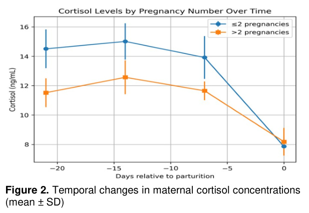
When comparing cortisol levels on days 21, 14, and 7 before birth and the postnatal period (day 0) based on the number of lambs born, a statistically significant decrease in cortisol levels was observed on day 0 (p<0.05) (Table 1).When cortisol levels were examined according to the number of pregnancies in the sheep, no significant difference was found (p>0.05) (Table 2).
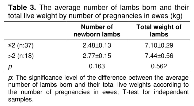
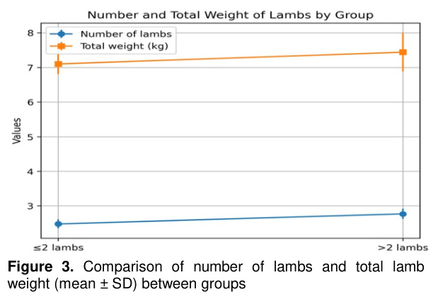
No significant difference was observed between the number of lambs born and their total average live weights based on the number of pregnancies in the ewes (p>0.05) (Table 3).
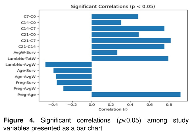
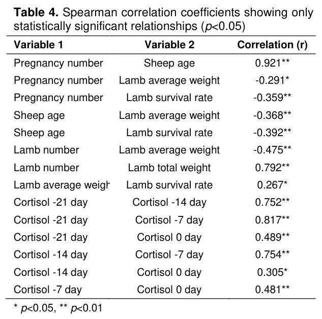
Table 4 presents the correlation results between pregnancy number and sheep age (0.921**); pregnancy number and lamb average weight (-0.291*); pregnancy number and lamb survival rate (-0.359**); sheep age and lamb average weight (-0.368**); sheep age and lamb survival rate (-0.392**); lamb number and lamb average weight (-0.475**); lamb number and lamb total weight (0.792**); lamb average weight and lamb survival rate (0.267*); cortisol on day -21 and cortisol on day -14 (0.752**); cortisol on day -21 and cortisol on day -7 (0.817**); cortisol on day -21 and cortisol on day 0 (0.489**); cortisol on day -14 and cortisol on day -7 (0.754**); cortisol on day -14 and cortisol on day 0 (0.305*); and cortisol on day -7 and cortisol on day 0 (0.481**) (* p<0.05, ** p<0.01).
The average live weights of singleton lambs were 3.4 kg, twin lambs 3.03 kg, triplet lambs 2.78 kg, and quadruplet lambs 2.24 kg. A total of 142 lambs were included in the survival analysis. During the first 24 hours after birth, 123 lambs survived and 19 lambs died, corresponding to a survival rate of 86.6% and a mortality rate of 13.4%.
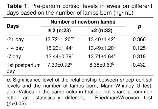
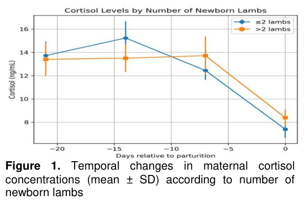
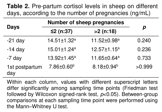
Within each column, values with different superscript letters differ significantly among sampling time points (Friedman test followed by Wilcoxon signed-rank test, p<0.05). Between-group comparisons at each sampling time point were performed using the Mann–Whitney U test.
Discussion
Glucocorticoids, such as cortisol, play a crucial role in regulating metabolism in both adult and fetal tissues. Circulating glucocorticoid concentrations typically increase during late gestation; however, exposure of the dam to stressors such as undernutrition, infection, or transportation may induce a premature elevation in cortisol levels [11]. In addition, multiple fetuses may further influence the increase in maternal cortisol concentrations during late gestation [2]. In a study conducted in sheep, cortisol concentrations were reported to reach peak levels approximately two weeks prior to parturition [12]. Similarly, a study in sheep demonstrated that cortisol concentrations were higher in twin-bearing ewes compared with those carrying a single fetus [4]. In a study conducted in Afec-Assaf ewes, maternal cortisol concentrations during late gestation were reported to differ between those carrying two or more fetuses and those bearing a single fetus [6]. Tharwat et al. [13] reported that cortisol concentrations reached their peak at parturition in goats during the transition period. Based on the findings of the present study, no differences were observed in cortisol concentrations between days −21, −14, and −7 among groups stratified according to litter size and parity in Romanov ewes. However, cortisol concentrations on day 0 were found to be significantly lower than those measured at the other time points (p<0.05). In the present study, the absence of marked changes in cortisol concentrations during late gestation—particularly in Romanov ewes carrying triplets and quadruplets—may indicate a greater resilience to stress in this breed.Maternal stress is widely recognized as one of the primary contributors to elevated fetal stress hormone levels and, in addition to adversely affecting fetal development, may predispose offspring to health disorders later in life [14]. Glucocorticoid administration in pregnant monkeys, sheep, and mice has been reported to result in impaired fetal growth [15]. Downs et al. [16] and Alon et al. [6] reported a negative association between maternal cortisol concentrations and lamb birth weights in studies conducted in Dall and Afec-Assaf sheep, respectively. In contrast, no negative association was observed between lamb birth weights and maternal cortisol concentrations in the present study. Further large-scale investigations are warranted to elucidate the underlying reasons for the absence of such a relationship between late-gestation cortisol levels and birth weight in Romanov ewes.
Another key determinant of fetal growth is the number of fetuses carried by the dam. In sheep, an increase in litter size is generally associated with a reduction in lamb birth weight [17]. Consistent with the findings of Miller et al. [18], the present study also demonstrated a negative relationship between litter size and the mean birth weights of lambs.
It is known that there is a relationship between lamb survival and growth rates and birth weight. Generally, smaller lambs have lower survival rates and daily weight gains compared to larger lambs [19]. Schreurs et al. [20] found a negative correlation between birth weight and mortality rates in twin lambs. Juengel et al. [19] reported that reduced body weight in lambs adversely affects both survival and developmental performance. Consistent with these findings, the present study demonstrated a negative association between lamb birth weight and mortality rate, which may be attributed to decreased disease resistance in lower-weight lambs. A possible explanation for the absence of an association between maternal cortisol concentrations and lamb birth weight is that cortisol concentrations measured during late gestation may not fully reflect cumulative fetal exposure to glucocorticoids throughout pregnancy. Furthermore, genetic characteristics of Romanov sheep, maternal nutritional status, placental efficiency, and other endocrine mechanisms may have exerted a stronger influence on fetal growth than maternal cortisol concentrations alone.
Aktaş et al. [21] reported that the age of the dam influences lamb survival rates. In their study on Central Anatolian Merino sheep, lambs born to ewes aged 3.5–4.5 years exhibited higher survival rates compared with those born to 1.5-year-old ewes. Sawalha et al. [22] reported that lambs born to young (1–2 years) and older (≥5 years) ewes exhibited lower survival rates compared with those born to middle-aged ewes (3–5 years) in Scottish Blackface sheep. Consistent with these findings, the present study demonstrated a negative association between maternal age and parity and lamb survival rates. The observed negative correlations between ewe age and lamb survival rate, as well as lamb average birth weight, may be related to differences in maternal physiological condition, body condition, and reproductive performance among ewes of different ages. These factors may influence fetal development during gestation and the lambs’ subsequent survival.
Conclusion
In conclusion, it is noteworthy that in highly prolific Romanov ewes, maternal cortisol concentrations during late gestation—a key stress hormone—appear to be unaffected by litter size and do not exert a significant influence on lamb survival rates. Further comprehensive and advanced studies are warranted to elucidate the underlying mechanisms of this finding. Moreover, considering these superior reproductive traits, Romanov sheep may be regarded as an economically advantageous breed for livestock producers. One limitation of the present study was the relatively small number of ewes carrying triplets and quadruplets, which prevented separate statistical evaluation of these groups. Additional metabolic and endocrine parameters potentially affecting lamb survival were not evaluated. Future studies including larger populations of highly prolific Romanov ewes are warranted to confirm the present findings. In addition, an a priori sample size (power) calculation was not performed, and the study population was limited to the available eligible animals in the research flock. Therefore, larger prospective studies with predefined sample size calculations are recommended to validate these findings.References
- Ibrahim MA, Abdelrahman H, Elmetwaly H. Hormonal profile, antioxidant status and some biochemical parameters during pregnancy and periparturient period in dromedary she camel. Egypt J Vet Sci 2017; 48: 81-94.
- Akbulut NK, Harman H, Kal Y, Kirbas M. Examination of blood cortisol and some parameters at parturition and on 30th day postpartum in single and twin - pregnant ewes. Livest Stud 2021; 61: 55-59.
- Walejko JM, Antolic A, Koelmel JP, et al. Chronic maternal cortisol excess during late gestation leads to metabolic alterations in the newborn heart. Am J Physiol Endocrinol Metab 2019; 316: 546-556.
- Medan M, Al-daek T, Absy G. Changes in serum cortisol level during pregnancy in ewes and the effect of fetal number. Suez Canal Vet Med J 2015; 20: 117-133.
- Zijlmans MAC, Riksen-Walraven JM, Weerth C. Associations between maternal prenatal cortisol concentrations and child outcomes: A systematic review. Neurosci Biobehav Rev 2015; 53: 1-24.
- Alon T, Matas D, Koren L, Gootwine E. Higher cortisol and testosterone levels in sheep with larger litter sizes. Livest Sci 2021; 243: 1-5.
- Vostry L, Milerski M, Schmidova J, Vostra-Vydrova H. Genetic diversity and effect of inbreeding on litter size of the Romanov sheep. Small Ruminant Res 2018; 168: 25-31.
- Aziz NM, Dvalishvili VG. Growth performance and carcass quality assessment of purebred and crossbred romanov lambs. Mesop J Agric 2020; 48: 35-42.
- Ricordeau G, Thimonier J, Poivey JP, et al. I.N.R.A. research on the romanov sheep breed in france: A review. Livest Prod Sci 1990; 24: 305-332.
- Dvalishvili VG, Fathala MM, Vinogradov IS, Dawod A. Influence of crossbreeding Romanov ewes with crossbred Argali Romanov rams on male progeny performance and carcass traits. J Vet Sci Technol 2015; 6: 1-6.
- Vaughan OR, Davies KL, Ward JW, Blasio MJ, Fowden AL. A physiological increase in maternal cortisol alters uteroplacental metabolism in the pregnant ewe. J Physiol 2016; 594: 6407–6418.
- Greguła-Kania M, Kosior-Korzecka U, Patkowski K, et al. Acute-phase proteins, cortisol and haematological parameters in ewes during the periparturient period. Reprod Domest Anim 2020; 55: 393-400.
- Tharwat M, Ali A, Al-Sobayil F. Hematological and biochemical profiles in goats during the transition period. Comp Clin Pathol 2015; 24:1-7.
- Hermesa M, Antonow-Schlorkea I, Hollsteina D, et al. Maternal psychosocial stress during early gestation impairs fetal structural brain development in sheep. Stress 2020; 23: 233-242.
- Jensen EC, Gallaher BW, Breier BH, Harding JE. The effect of a chronic maternal cortisol infusion on the late-gestation fetal sheep. J Endocrinol 2002; 174: 27-36.
- Downs DJ, Boan BV, Lohuis TD, Stewart KM. Investigating relationships between reproduction, immune defenses, and cortisol in Dall sheep. Front Immunol 2018; 9: 1-11.
- Gootwine E. Variability in the rate of decline in birth weight as litter size increases in Sheep. Anim Sci 2005; 81: 393-398.
- Miller DR, Blache D, Jackson RB, Downie EF, Roche JR. Metabolic maturity at birth and neonate lamb survival: Association among maternal factors, litter size, lamb birth weight, and plasma metabolic and endocrine factors on survival and behavior. J Anim Sci 2013; 88: 581–592.
- Juengel JL, Davisa GH, Wheelerb R, Doddsa KG, Johnstonea PD. Factors affecting differences between birth weight of littermates (BWTD) and the effects of BWTD on lamb performance. Anim Reprod Sci 2018; 191: 34-43.
- Schreurs NM, Kenyon PR, Mulvaney FJ, et al. Effect of birthweight and birth rank on the survival of single and twin lambs born to ewe lambs. Anim Prod Sci 2010; 50: 460-464.
- Aktaş AH, Dursun Ş, Doğan Ş, et al. Effects of ewe live weight and age on reproductive performance, lamb growth, and survival in Central Anatolian Merino sheep. Arch Anim Breed 2015; 58: 451-459.
- Sawalha RM, Conington J, Brotherstone S, Villanueva B. Analyses of lamb survival of Scottish Blackface sheep. Animal 2007; 1: 151-157.
- KUZUDA RASTLANILAN MONOCEPHALIEN OLGUSU
- PAZARLANAMAYAN KIRIK YUMURTALARIN TOKLU BESİ RASYONLARINDA KULLANILMA OLANAKLARI
- KUZULARDA GÖZYAŞI SEKRESYONUNUN İPLİK TESTİ İLE KLİNİK OLARAK DEĞERLENDİRİLMESİ
- YENİ DOĞAN KUZULARDA ARTERİYEL OKSİJEN SATURASYONUNUN PULSE OKSİMETRE İLE BELİRLENMESİ
- ARPA FİĞ HASILININ EN UYGUN BALYALAMA ZAMANININ BELİRLENMESİ VE KUZU BESİSİNDE SAMAN YERİNE FARKLI PROTEİN KAYNAKLARI İLE KULLANILMASI*