
Barınak Köpeklerinde Plazma Kortizol Konsantrasyonları ile Hematolojik Profil Arasındaki İlişki
Volume 40, Issue 3
October 2026 · Pages 255-260
Barınak Köpeklerinde Plazma Kortizol Konsantrasyonları ile Hematolojik Profil Arasındaki İlişki
Relationship Between Plasma Cortisol Concentrations and Hematological Profile in Shelter Dogs
Elmas ULUTAŞ1, Çağatay SALUM2, Özlem KATMER3
2Kastamonu University, Faculty of Veterinary Medicine, Department of Physiology, Kastamonu, TÜRKİYE
3Yozgat Bozok University, Faculty of Veterinary Medicine, Undergraduate Student, Yozgat, TÜRKİYE
Bu çalışmada, aynı barınakta yaşayan klinik olarak sağlıklı 66 erişkin erkek melez köpekte plazma kortizol düzeyleri ile hematolojik parametreler arasındaki ilişkinin belirlenmesi amaçlandı. Kanlarda tam kan sayımı yapıldı ve plazma kortizol konsantrasyonları ELISA kiti kullanılarak ölçüldü. Örnekler kortizol düzeylerine göre düşük, orta ve yüksek olmak üzere üç eşit gruba ayrıldı. Düşük kortizol grubuyla karşılaştırıldığında, yüksek kortizol grubunda lökosit, nötrofil ve monosit sayıları daha yüksek, lenfosit sayısı ise daha düşüktü. Düşük kortizol grubuna kıyasla yüksek kortizol grubunda lenfosit yüzdesi daha düşüktü; monosit yüzdesi ise orta kortizol grubunda diğer iki gruptan daha yüksekti. Diğer lökosit parametreleri gruplar arasında farklılık göstermedi. Eritrosit parametrelerinden ortalama eritrosit hacmi, ortalama eritrosit hemoglobini ve eritrosit dağılım genişliği-varyasyon katsayısı kortizol grupları arasında farklılık gösterirken, diğer eritrosit parametreleri benzerdi. Trombosit parametreleri açısından ise yalnızca ortalama trombosit hacmi kortizol grupları arasında farklılık gösterdi. Korelasyon analizinde plazma kortizol konsantrasyonlarının lökosit, nötrofil ve monosit sayıları, monosit yüzdesi, ortalama eritrosit hacmi ve ortalama eritrosit hemoglobini ile anlamlı pozitif korelasyon gösterdiği belirlendi. Buna karşılık, lenfosit yüzdesi ve nötrofil/lenfosit oranı ile anlamlı negatif korelasyonlar saptandı. Sonuç olarak, plazma kortizol konsantrasyonları ile lökosit parametreleri arasındaki ilişkiler daha belirginken, eritrosit ve trombosit parametreleriyle olan ilişkiler daha sınırlıydı. Bu sonuçlar, plazma kortizol konsantrasyonları ile hematolojik parametrelerin birlikte ele alınmasının, barınaktaki köpeklerde fizyolojik stres yanıtının daha kapsamlı yorumlanmasına katkı sağlayabileceğini düşündürmektedir.
The aim of this study was to determine the relationship between plasma cortisol levels and hematological parameters in 66 clinically healthy adult male mixed-breed dogs housed in the same shelter. A complete blood count was performed, and cortisol concentrations were measured using an ELISA kit. The samples were divided into three equal groups based on their cortisol levels: low, medium, and high. In comparison with the low cortisol group, the high cortisol group had higher white blood cell, neutrophil, and monocyte counts, while the lymphocyte count was lower. Compared with the low group, lymphocyte percentage was lower in the high group; monocyte percentage was higher in the medium than in both other groups. Other leukocyte parameters did not differ. Among the erythrocyte parameters, mean corpuscular volume, mean corpuscular hemoglobin, and erythrocyte distribution width (coefficient of variation) differed among the cortisol groups, while the other erythrocyte parameters were similar. For platelet parameters, the only difference observed among the cortisol groups was in the mean platelet volume. In the correlation analysis, it was determined that cortisol concentrations were significantly positively correlated with leukocyte, neutrophil, and monocyte counts, monocyte percentage, mean corpuscular volume, and mean corpuscular hemoglobin. In contrast, plasma cortisol concentrations were significantly negatively correlated with lymphocyte percentage and the neutrophil-to-lymphocyte ratio. In summary, while cortisol relationships with leukocyte parameters were more evident, those with erythrocyte and platelet parameters were limited. The results suggest that evaluating plasma cortisol concentrations and hematological parameters together may provide a more comprehensive interpretation of physiological stress response in shelter dogs.
Introduction
In dogs living in shelters, evaluating animal welfare poses a significant challenge as physiological and behavioral responses are influenced by numerous environmental and individual factors [1]. Conditions in the shelter, such as limited social interaction, reduced environmental control, unpredictable noise levels, and variations in care routines, can affect the physiological status and welfare of dogs [1,2]. In addition, restricted regular interaction with people can lead to desocialization in dogs, thereby weakening their ability to deal with stressors [3]. Individual differences in adaptive capacity make it even more difficult to objectively assess welfare status [2]. For this purpose, interest in the evaluation of the physiological status of dogs living in shelters using reliable biomarkers has increased in recent years [1].One of the most commonly used physiological biomarkers in the monitoring of animal welfare is cortisol. Because it can be measured in a wide variety of biological samples - such as blood (plasma/serum), saliva, urine, feces, and hair - and reflects the activation of the hypothalamic-pituitary-adrenal (HPA) axis, it is used as a standard means of assessing the stress response in veterinary research [1,4]. However, since cortisol levels are subject to fluctuations due to physiological, environmental, and individual variables such as age, body weight, breed, sex, and circadian rhythm, they may not be sufficient on their own to comprehensively understand animal welfare or stress levels [4,5]. Holistic approaches based on the interpretation of cortisol in conjunction with other physiological biomarkers are increasingly gaining acceptance [4].
The physiological stress response is a complicated process involving the HPA axis as well as the interactions among the nervous, endocrine, immune, and cardiovascular systems. Assessing the stress response only through cortisol levels may not be sufficient to reveal all the physiological alterations occurring in the body [4,6,7]. The changes that occur in leukocyte subpopulations - particularly neutrophilia, lymphopenia, and the resulting changes in the neutrophil-to-lymphocyte (N/L) ratio - are considered important hematological indicators of the physiological stress response [8,9]. In recent years, it has been reported that the N/L ratio is an accessible hematological biomarker that reflects the balance between innate and adaptive immune responses, with cortisol and catecholamines as its main determinants [10].
A complete blood count is one of the fundamental laboratory tests widely used in veterinary clinical practice that allows for the simultaneous determination of leukocyte, erythrocyte, and thrombocyte parameters from the same blood sample [8,11]. Since complete blood count analysis is rapid, inexpensive, and routinely performed in veterinary practice, hematological parameters have the potential to serve as practical and complementary biomarkers in the assessment of physiological stress. Therefore, identifying hematological changes associated with different plasma cortisol concentrations may contribute to a more comprehensive assessment of stress and welfare in shelter dogs. The interpretation of cortisol levels in conjunction with complete blood count results contributes to a more comprehensive assessment of the physiological stress response [4]. It is known that the shelter environment is one of the key environmental factors influencing the long-term physiological stress response in dogs [2]. However, studies examining the relationship between plasma cortisol levels and the hematological profile in clinically healthy dogs living in shelters are limited [12,13]. The aim of this study was to determine the association between plasma cortisol levels and hematological parameters obtained from a complete blood count in clinically healthy adult male dogs housed in the same shelter.
Material and Method
The study population consisted of 66 adult male mixed-breed dogs, aged 1-3 years and weighing 25-45 kg, housed at the Sorgun Municipality Animal Shelter under identical care and feeding conditions. The dogs were enrolled in the study during routine clinical examinations conducted at the shelter. Only clinically healthy dogs that showed no signs of systemic disease and were not receiving any treatment were included in the study. To minimize the effects of circadian variation in plasma cortisol concentrations, blood samples were collected by cephalic vein (vena cephalica) between 08:30 and 10:30 into dipotassium ethylenediaminetetraacetic acid (K2EDTA) containing tubes.The complete blood count (CBC) tests were carried out on a hemogram analyzer (Mindray BC-5000; Shenzhen Mindray Bio-Medical Co., Ltd., China) as soon as possible after blood samples were collected.
After hematological analysis, blood samples were centrifuged at 2000 rpm for 15 min at 4°C using a refrigerated centrifuge (Universal 320R; Andreas Hettich GmbH & Co. KG, Tuttlingen, Germany). The plasma samples were stored in a -80°C deep-freeze (ESCO ESC-UUS-480A, ESCO, Singapore) until cortisol analyses were conducted.
Plasma cortisol concentrations were measured using a General Cortisol (COR) ELISA kit (BT Laboratory, Shanghai, China; Cat. No. EA0010Ge) according to the manufacturer’s instructions. The assay had a detection range of 0.5–300 ng/mL, a sensitivity of 0.27 ng/mL, an intra-assay coefficient of variation (CV) of <10%, and an inter-assay CV of <12%. Absorbance values were measured at 450 nm using an ELISA microplate reader (SPECTROstar Nano; BMG LABTECH, Offenburg, Germany).
This study was reviewed by the Local Ethics Committee for Animal Experiments at Yozgat Bozok University (Meeting Date: May 28, 2025; Meeting No.: 2025/4; Decision No.: 7). The study was conducted with permission from the Sorgun Municipality Animal Shelter Directorate. Blood samples were collected during routine veterinary clinical work. No additional invasive or experimental procedures were performed on the animals for research purposes.
The sample size to be used in the study was calculated using power analysis with the G*Power 3.1 software. Since the analyses were conducted with three groups, the “ANOVA: Fixed effects, omnibus, one-way” model was used. The calculations used a Cohen effect size (f) of 0.40, a significance level (α) of 0.05, a test power (1-β) of 0.80. Similarly, in a study investigating the accumulation of toxic elements in dogs, it was stated that the selected parameters were sufficient for statistical analysis [14]. The analysis found that the required number of animals for each group was 22. The total number of animals was also calculated to be 66. Cortisol concentrations were ranked from lowest to highest, and the 66 dogs were divided into three equal-sized groups; low, medium and high. This classification was based on the relative distribution of cortisol concentrations within the dog population.
Statistical analyses of the data were performed using the SPSS 23.0 software program (SPSS Inc., Chicago, IL, USA). The homogeneity of variances was checked using Levene’s test. The normality of the data was assessed using the Shapiro-Wilk test. The significance of the data for parameters following a normal distribution was examined using One-way ANOVA. Differences between groups for these parameters were determined using the Tukey post-hoc test. Parameters that did not follow normal distribution were analyzed using the Kruskal-Wallis test. The Dunn-Bonferroni multiple comparison test was applied to the variables for which significant differences were detected. The relationships between plasma cortisol concentrations and hematological parameters were evaluated using Spearman’s rank correlation analysis, as several hematological variables did not meet the assumption of normality. Cortisol concentration was included in the correlation analysis as a continuous variable. Spearman’s rank correlation coefficient (ρ) and corresponding p values were reported. All analyses were considered significant at the 95% confidence level (p<0.05).
Results
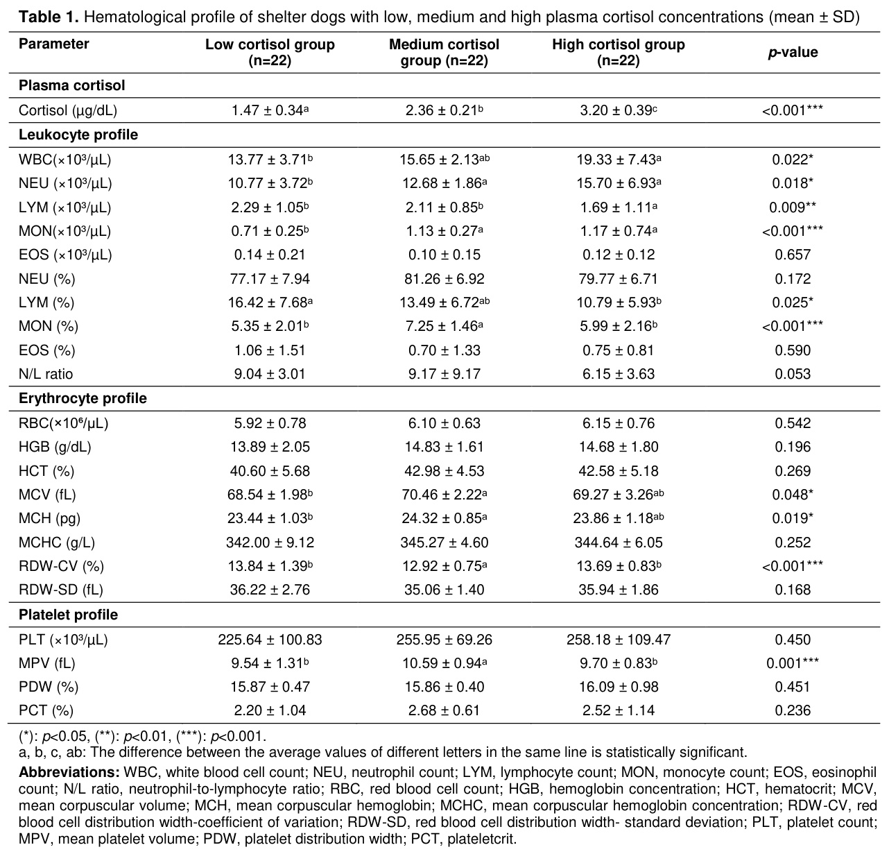
Plasma cortisol concentrations exhibited statistically significant differences among the low, medium, and high cortisol groups (p<0.001) (Table 1) a, b, c, ab: The difference between the average values of different letters in the same line is statistically significant.
Abbreviations: WBC, white blood cell count; NEU, neutrophil count; LYM, lymphocyte count; MON, monocyte count; EOS, eosinophil count; N/L ratio, neutrophil-to-lymphocyte ratio; RBC, red blood cell count; HGB, hemoglobin concentration; HCT, hematocrit; MCV, mean corpuscular volume; MCH, mean corpuscular hemoglobin; MCHC, mean corpuscular hemoglobin concentration; RDW-CV, red blood cell distribution width-coefficient of variation; RDW-SD, red blood cell distribution width- standard deviation; PLT, platelet count; MPV, mean platelet volume; PDW, platelet distribution width; PCT, plateletcrit.
Analysis of the leukocyte profile revealed significant differences in WBC, NEU, LYM, and MON counts, as well as LYM% and MON%, across the cortisol groups. The differences were significant at p<0.05 for WBC, NEU, and LYM%, at p<0.01 for LYM, and at p<0.001 for MON and MON%. Post hoc analysis showed that WBC, NEU, and MON counts were higher, whereas the LYM count was lower, in the high cortisol group than in the low cortisol group. Furthermore, MON% was higher in the medium cortisol group than in both the low and high cortisol groups, whereas LYM% was lower in the high cortisol group than in the low cortisol group. No significant differences were observed for EOS count, NEU%, EOS%, or the N/L ratio (p>0.05).
Among the erythrocyte parameters, MCV and MCH differed significantly among the cortisol groups (p<0.05). For both parameters, values in the medium cortisol group were higher than those in the low cortisol group, whereas the high cortisol group did not differ significantly from either group. RDW-CV also differed significantly among the groups (p<0.001) and was lower in the medium cortisol group than in the low and high cortisol groups. No significant differences were observed for RBC, HGB, HCT, MCHC, or RDW-SD (p>0.05).
The platelet parameters were examined, and only the Mean Platelet Volume (MPV) showed a significant difference among the cortisol groups (p=0.001). Post hoc analysis showed that MPV was higher in the medium cortisol group than in the low and high cortisol groups. No significant differences were observed for PLT, PDW, or PCT (p>0.05).
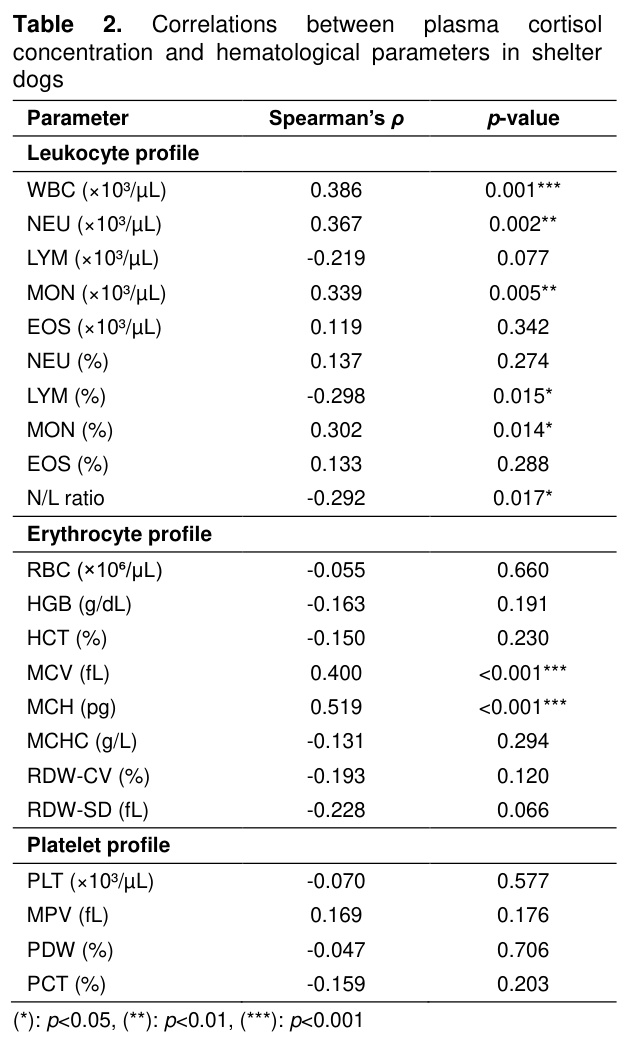
Correlation analysis (Table 2) revealed significant positive correlations between plasma cortisol concentrations and WBC (ρ=0.386, p=0.001), NEU (ρ=0.367, p=0.002), MON (ρ=0.339, p=0.005), MON (%) (ρ=0.302, p=0.014), MCV (ρ=0.400, p<0.001), and MCH (ρ=0.519, p<0.001). Significant negative correlations were observed between plasma cortisol concentrations and LYM (%) (ρ=-0.298, p=0.015) and the N/L ratio (ρ=-0.292, p=0.017). No significant correlations were found between plasma cortisol concentrations and LYM, EOS, NEU (%), EOS (%), RBC, HGB, HCT, MCHC, RDW-CV, RDW-SD, PLT, MPV, PDW, or PCT (p>0.05).
Discussion
In this study, both group comparisons and correlation analyses demonstrated that plasma cortisol concentrations in clinically healthy male shelter dogs were associated with selected hematological parameters. While significant differences were observed, particularly in leukocyte parameters, among groups formed based on cortisol levels, the correlation analysis revealed that, in addition to several leukocyte parameters, MCV and MCH values also demonstrated a significant relationship with plasma cortisol concentrations. In contrast, the majority of erythrocyte and platelet parameters did not exhibit significant differences among the cortisol groups nor did they reveal significant correlations with plasma cortisol concentrations.Although cortisol is one of the most commonly used biomarkers for assessing the physiological stress response, plasma cortisol concentrations can be influenced by several factors, including sampling time, environmental conditions, individual variability, and adaptation to the environment. Instead of evaluating cortisol alone, current approaches recommend interpreting it in conjunction with other physiological and behavioral indicators [4,5].
In our study, dogs with high plasma cortisol concentrations had higher WBC, NEU, and MON counts and lower LYM counts than dogs with low cortisol concentrations. In addition, correlation analysis revealed positive correlations between plasma cortisol concentrations and WBC, NEU, and MON counts, as well as MON%, while LYM% correlated negatively with plasma cortisol concentration. The leukocyte changes observed in the present study are generally consistent with the glucocorticoid-associated leukogram described in dogs, which is characterized by neutrophilia, lymphopenia, and, in some cases, monocytosis [8,9,11]. Consistent with these findings, Dudley et al. [15] reported higher total leukocyte, neutrophil, and monocyte counts in shelter dogs than in control dogs. In contrast, Tüfekçi et al. [16] reported a gradual decrease in cortisol concentrations in hospitalized dogs; however, no significant changes were detected in most hematological parameters during the observation period.
Although NEU counts were higher and LYM counts were lower in the high cortisol group, the N/L ratio did not differ significantly among the cortisol groups. However, correlation analysis revealed a statistically significant negative correlation between plasma cortisol concentrations and the N/L ratio. These apparently discordant results may reflect differences in the temporal dynamics of plasma cortisol and leukocyte responses to stress, as these measures do not necessarily change in parallel. Davis and Maney [17] emphasized that glucocorticoid concentrations and N/L ratios are not interchangeable stress indicators because the hormonal response generally develops and resolves more rapidly than the leukocyte response. Similarly, Yun et al. [18] reported that the N/L ratio in dogs with hypercortisolism was higher than in healthy dogs and dogs with non-adrenal diseases, but that there was no significant correlation between the N/L ratio and cortisol concentrations measured before or after ACTH administration. Squair et al. [19] also observed a decrease in serum cortisol concentrations during repeated veterinary visits but did not determine a corresponding significant change in the N/L ratio. Likewise, Dudley et al. [15] reported that plasma cortisol concentrations decreased during the shelter housing, while the N/L ratio remained high. When considered together, these results suggest that plasma cortisol concentrations and the N/L ratio may be related to different temporal components of the stress response and may therefore follow different patterns over time.
Previous studies investigating the effects of cortisol and the physiological stress response on erythrocyte parameters have reported inconsistent findings. In hospitalized dogs, marked changes in cortisol concentrations were accompanied by significant differences in HGB, MCV, and MCH at certain measurement times, whereas RBC, HCT, and RDW did not differ significantly [16]. Similarly, Karić et al. [20] reported no significant differences in RBC, HGB, HCT, MCV, MCH, or MCHC between dogs with and without hyperadrenocorticism, whereas PLT was the only hematological parameter that differed significantly. Studies using different stress models have also reported variable results for erythrocyte parameters [21-23]. In the present study, erythrocyte count, hemoglobin concentration, hematocrit, MCHC, and RDW-SD did not differ among the cortisol groups. Significant differences were observed for MCV, MCH, and RDW-CV; however, these parameters did not exhibit a consistent increase or decrease across the cortisol groups. Correlation analysis also revealed significant positive correlations between plasma cortisol concentrations and MCV and MCH, whereas no significant correlation was observed for RDW-CV. MCV, MCH, and RDW provide information on erythrocyte size, hemoglobin content, and variation in erythrocyte volume, respectively, and may vary with changes in erythropoiesis and iron availability [8,11]. Glucocorticoids regulate erythroid progenitor cell proliferation and contribute to stress erythropoiesis, particularly under conditions of increased erythropoietic demand [24,25]. Therefore, the differences observed in MCV, MCH, and RDW-CV, together with the positive correlations of MCV and MCH with plasma cortisol concentrations, should be interpreted cautiously, as these erythrocyte indices may be associated with multiple physiological processes. In addition, unmeasured factors such as individual differences in hydration status or undetected subclinical conditions may have contributed to the variability observed in these hematological parameters.
Among the platelet parameters, only MPV differed among the cortisol groups, whereas PLT, PCT, and PDW did not differ significantly. MPV represents the average size of circulating platelets, and increased MPV may be associated with enhanced platelet production and the presence of younger platelets [8,11]. Experimental studies indicate that glucocorticoids may modulate platelet production and function through their effects on megakaryocytes and platelet signaling pathways [26-28]. However, Tüfekçi et al. [16] reported no significant differences in MPV or PLT despite significant changes in serum cortisol concentrations in hospitalized dogs. Similarly, Kulka et al. [13] reported no significant differences in platelet count or other routine hematological parameters among short-term shelter, long-term shelter, and client-owned dogs. In the present study, although MPV differed among the cortisol groups, no significant correlation was observed between plasma cortisol concentrations and MPV. In contrast, PLT, PCT, and PDW did not differ significantly among the cortisol groups, and none of these parameters significantly correlated with plasma cortisol concentrations. Therefore, the isolated difference in MPV should not be interpreted as direct evidence of platelet activation. Further studies including specific markers of platelet activation are needed to better clarify the relationship between cortisol concentrations and platelet function.
This study demonstrated that variations in plasma cortisol concentrations are not uniformly associated with hematological parameters in clinically healthy shelter dogs. Associations with plasma cortisol concentrations were more evident for leukocyte parameters, whereas those involving erythrocyte and platelet parameters were more limited. These findings suggest that hematological profiling, when evaluated alongside plasma cortisol concentrations, may provide complementary information for the assessment of the physiological stress response in shelter dogs.
In practical terms, the combined evaluation of plasma cortisol concentrations and hematological profiles may assist veterinarians and shelter personnel in the assessment of physiological stress and animal welfare, particularly when interpreted together rather than as isolated indicators. Nevertheless, plasma cortisol reflects relatively short-term changes in HPA axis activity, whereas shelter living may represent a longer-term stress condition. Therefore, the absence of hair cortisol measurements, which may provide information on cumulative cortisol exposure over a longer period, should be considered a limitation of the present study. In addition, hydration status and potential subclinical conditions, including parasitic infections, were not specifically evaluated and may have contributed to individual variation in hematological parameters. These limitations should be considered when interpreting the differences observed in MCV, MCH, and MPV. Future studies combining plasma and hair cortisol measurements with a more comprehensive assessment of factors that may influence hematological parameters may help clarify the biological significance of cortisol-associated hematological changes.
References
- Righi C, Menchetti L, Orlandi R, et al. Welfare assessment in shelter dogs by using physiological and immunological parameters. Animals 2019; 9: 340.
- Protopopova A. Effects of sheltering on physiology, immune function, behavior, and the welfare of dogs. Physiol Behav 2016; 159: 95-103.
- Gazzano V, Curadi MC, Baragli P, et al. Physiological and behavioral evaluation of shelter dogs during veterinary routine health checks. Vet Sci 2025; 12: 583.
- Cobb ML, Jimenez AG, Dreschel NA. Beyond cortisol! Physiological indicators of welfare for dogs: Deficits, misunderstandings and opportunities. J Appl Anim Welf Sci 2026; 29: 951-980.
- Kaszycka K, Goleman M, Krupa W. Testing the level of cortisol in dogs. Animals 2025; 15: 1197.
- McEwen BS. What is the confusion with cortisol? Chronic Stress 2019; 3: 1-3.
- Oken BS, Chamine I, Wakeland W. A systems approach to stress, stressors and resilience in humans. Behav Brain Res 2015; 282: 144-154.
- McCourt MR, Rizzi TE. Hematology of dogs. In: Brooks Marjory B, Harr KE, Seelig DM, Wardrop KJ, Weiss DJ (Editors). Schalm’s Veterinary Hematology. 7th Edition, Philadelphia: John Wiley & Sons, Inc 2022: 969-982.
- Stockham SL, Keeton KS, Szladovits B. Clinical assessment of leukocytosis: Distinguishing leukocytoses caused by inflammatory, glucocorticoid, physiologic, and leukemic disorders or conditions. Vet Clin Small Anim Pract 2003; 33: 1335-1357.
- Muñoz AI, Maldonado-García JL, Fragozo A, et al. Altered neutrophil-to-lymphocyte ratio in sepsis secondary to canine parvoviral enteritis treated with and without an immunomodulator in puppies. Front Vet Sci 2022; 9, 995443.
- Stockham SL, Scott MA. Fundamentals of Veterinary Clinical Pathology. 2nd Edition, Ames: Blackwell Pub, 2008.
- Kulka M, Szopa IM, Klockiewicz M. Hematological changes and immunomodulation of neutrophil and monocyte populations in shelter dogs. Animals 2025; 15, 13: 1988.
- Kulka M, Szopa IM, Mizera-Szpilka K, et al. Stress-related immunomodulation of canine lymphocyte responses and hematologic profiles. Int J Mol Sci 2026; 27, 3: 1506.
- Bruno F, Miller A, Bruschetta G, et al. Levels of mineral elements in different organs of dogs from the ionian-etnean volcanic area. Animals 2025; 15: 1545.
- Dudley ES, Schiml PA, Hennessy MB. Effects of repeated petting sessions on leukocyte counts, intestinal parasite prevalence, and plasma cortisol concentration of dogs housed in a county animal shelter. J Am Vet Med Assoc 2015; 247: 1289-1298.
- Tüfekçi E, Ekinci G, Karaca Bekdik İ, et al. Determination of serum cortisol levels as a stress marker in hospitalized dogs. Turkish J Vet Anim Sci 2025; 49, 6: 288-298.
- Davis AK, Maney DL. The use of glucocorticoid hormones or leucocyte profiles to measure stress in vertebrates: What’s the difference? Methods Ecol Evol 2018; 9: 1556-1568.
- Yun S, Yun T, Cha S, et al. Can neutrophil-to-lymphocyte and platelet-to-lymphocyte ratios be used as markers for hypercortisolism in dogs? Top Companion Anim Med 2024; 61: 100890.
- Squair C, Proudfoot K, Montelpare W, et al. Effects of changing veterinary handling techniques on canine behaviour and physiology part 1: Physiological measurements. Animals 2023; 13, 7: 1253.
- Karić E, Hodžić A, Zahirović A, et al. Biochemical and haematological parameters in dogs with Cushing’s syndrome. Vet Stanica 2021; 52: 413-419.
- Angle CT, Wakshlag JJ, Gillette RL, et al. Hematologic, serum biochemical, and cortisol changes associated with anticipation of exercise and short duration high-intensity exercise in sled dogs. Vet Clin Pathol 2009; 38: 370-374.
- Azeez OM, Olaifa FH, Adah AS, et al. Effect of heat stress on vital and hematobiochemical parameters of healthy dogs. Vet World 2022; 15: 722-727.
- Asogwa CN, Onyeka CC, Odii EC, et al. Haematological and cortisol dynamics in albino rats stimulated by intake of methanolic extract of cola acuminata. World J Med Sci 2014; 10: 384-391.
- Bauer A, Tronche F, Wessely O, et al. The glucocorticoid receptor is required for stress erythropoiesis. Genes Dev 1999; 13: 2996-3002.
- Hanssen LLP, Iskander D. The role of glucocorticoids in erythropoiesis. Front Hematol 2025; 4:1540152.
- Grodzielski M, Cidlowski JA. Glucocorticoids regulate thrombopoiesis by remodeling the megakaryocyte transcriptome. J Thromb Haemost 2023; 21: 3207-3223.
- Signorello MG, Ravera S, Leoncini G. Oxidative stress induced by cortisol in human platelets. Int J Mol Sci 2024; 25: 3776.
- Liverani E, Banerjee S, Roberts W, et al. Prednisolone exerts exquisite inhibitory properties on platelet functions. Biochem Pharmacol 2012; 83: 1364-1373.
- ELAZIĞ’DA KÖPEKLERDE TESPİT EDİLEN TRAVMATİK MYİASİSLER
- STUMP PYOMETRALI BİR KÖPEKTE OVARİAN REMNANT SENDROMU
- KÖPEKLERDE DENEYSEL TİBİA DEFEKTLERİNİN ONARIMINDA SİRKÜLER EKSTERNAL FİKSATÖRÜ KULLANILARAK YAPILAN KEMİK KAYDIRMA TEKNİĞİ UYGULAMASI
- KÖPEKLERDE UCU YİVLİ VE UCU YİVSİZ İNTRAMEDULLAR PİNLERİN AKSİYEL EKSTRAKSİYON GÜÇLERİNİN DENEYSEL OLARAK ARAŞTIRILMASI: HİSTOLOJİK BULGULAR
- BİR KÖPEKTE RASTLANAN TUBEROSİTAS TIBİAE’NİN AVULSİON KIRIĞI OLGUSU