
Tarçın, Streptozotosin ile Diyabet Oluşturulmuş Sıçanların Mide Dokusundaki Notch1-Pozitif Hücrelerin ve Metil Yeşili-Pironin-Pozitif Mast Hücrelerinin Sayısını Modüle Eder
Volume 40, Issue 3
October 2026 · Pages 288-296
Tarçın, Streptozotosin ile Diyabet Oluşturulmuş Sıçanların Mide Dokusundaki Notch1-Pozitif Hücrelerin ve Metil Yeşili-Pironin-Pozitif Mast Hücrelerinin Sayısını Modüle Eder
Cinnamon Modulates the Numbers of Notch1-Positive Cells and Methyl Green-Pyronin-Positive Mast Cells in the Gastric Tissue of Streptozotocin-Induced Diabetic Rats
Nazife ÜLKER ERTUĞRUL1, Tuğrul ERTUĞRUL2, Ahmet YARDIMCI3
2Ondokuz Mayıs University, Faculty of Veterinary Medicine, Department of Histology and Embryology, Samsun, TÜRKİYE
3Fırat University, Faculty of Medicine, Department of Physiology, Elazığ, TÜRKİYE
Diyabet mellitus, mide dokusunun hücresel bileşimini değiştirebilir ve mide dokusu homeostazında rol oynayan mekanizmaları etkileyebilir. Tarçın, antioksidan özelliklere sahip biyoaktif bileşikler içermekte olup bu bileşikler diyabetle ilişkili doku ve hücresel değişikliklerin iyileştirilmesine katkıda bulunabilir. Bu çalışma, streptozotosin (STZ) ile indüklenmiş diyabetik sıçanların mide dokusundaki Notch1-pozitif hücreler ve Metil Green-Pyronin (MGP)-pozitif mast hücre sayısına tarçın tedavisinin etkilerini araştırmayı amaçlamıştır. Otuz iki erkek sıçan rastgele dört gruba ayrıldı: kontrol, tarçın, diyabet ve diyabet+tarçın. Diyabet, tek bir intraperitoneal STZ enjeksiyonu (45 mg/kg) ile indüklendi ve tarçın ekstresi 14 gün boyunca 0.5 mg/kg dozunda oral olarak uygulandı. Notch1 pozitif hücreler immünohistokimyasal olarak değerlendirilirken, mast hücreleri MGP boyaması kullanılarak tanımlandı ve sayıldı. Hem Notch1-pozitif hücre sayısı hem de MGP-pozitif mast hücre sayısı için diyabet×tarçın etkileşimi anlamlı bulundu (p<0.0001). STZ ile indüklenen diyabet, kontrol grubuna kıyasla Notch1-pozitif hücre sayısını önemli ölçüde olarak azaltırken MGP-pozitif mast hücre sayısını artırdı (p<0.0001). Diyabetik olmayan koşullarda tarçın, Notch1-pozitif hücre sayısını anlamlı olarak değiştirmezken (p=0.660), MGP-pozitif mast hücre sayısını artırdı (p=0.044). Buna karşılık, diyabetik koşullarda tarçın tedavisi, tedavi edilmeyen diyabetik hayvanlara kıyasla Notch1-pozitif hücre sayısını anlamlı olarak artırırken MGP-pozitif mast hücre sayısını azalttı (p<0.0001). Tarçın, her iki parametrede de diyabetle ilişkili değişiklikleri hafifletmiş; Notch1-pozitif hücre sayısını kısmen eski haline getirirken, MGP-pozitif mast hücresi sayısını ise tarçın grubunda gözlemlenen seviyeye yakın bir düzeye düşürmüştür. Bu bulgular, tarçının mide dokusundaki Notch1-pozitif hücre ve MGP-pozitif mast hücre sayıları üzerindeki etkilerinin diyabetik duruma bağlı olduğunu ve diyabetik koşullarda her iki hücresel parametredeki diyabetle ilişkili değişiklikleri azalttığını düşündürmektedir.
Diabetes mellitus can alter the cellular composition of the gastric tissue and may affect mechanisms involved in its homeostasis. Cinnamon contains bioactive compounds with antioxidant properties that may contribute to ameliorating diabetes-associated tissue and cellular alterations. This study aimed to investigate the effects of cinnamon treatment on Notch1-positive cells and Methyl Green-Pyronin (MGP)-positive mast cell numbers in the gastric tissue of streptozotocin (STZ)-induced diabetic rats. Thirty-two male rats were randomly divided into four groups: control, cinnamon, diabetes, and diabetes+cinnamon. Diabetes was induced by a single intraperitoneal injection of STZ (45 mg/kg), and cinnamon extract was administered orally at 0.5 mg/kg for 14 days. Notch1-positive cells were evaluated immunohistochemically, whereas mast cells were identified and counted using MGP staining. Diabetes×cinnamon interactions were significant for both Notch1-positive cell number and MGP-positive mast cell number (p<0.0001). Diabetes significantly decreased Notch1-positive cell number and increased MGP-positive mast cell number compared with control animals (p<0.0001). Under non-diabetic conditions, cinnamon did not significantly alter Notch1-positive cell number (p=0.660) but increased MGP-positive mast cell number (p=0.044). Conversely, under diabetic conditions, cinnamon significantly increased Notch1-positive cell number and reduced MGP-positive mast cell number compared with untreated diabetic animals (p<0.0001). Cinnamon attenuated the diabetes-associated alterations in both parameters, partially restoring Notch1-positive cell number and reducing MGP-positive mast cell number to a level comparable with that observed in the cinnamon group. The effects of cinnamon on gastric Notch1-positive and MGP-positive mast cell numbers depend on diabetic status and that cinnamon attenuates diabetes-associated alterations in both cellular parameters under diabetic conditions.
Introduction
Diabetes mellitus (DM) is a chronic metabolic disorder characterized by persistent hyperglycemia resulting from impaired insulin secretion, impaired insulin action, or both [1]. Persistent hyperglycemia is a major contributor to the development of diabetic complications through mechanisms involving excessive reactive oxygen species production and oxidative stress [2]. Hyperglycemia-induced oxidative stress can promote cellular dysfunction and tissue injury through several interrelated metabolic pathways, including the polyol, hexosamine, protein kinase C, and advanced glycation end-product pathways [3]. In addition to its well-established vascular and neurological complications, diabetes can affect the gastrointestinal tract and lead to structural and functional abnormalities [4]. Long-standing diabetes is associated with gastric motor dysfunction and delayed gastric emptying, which are important manifestations of diabetic gastrointestinal involvement [5]. Experimental studies have demonstrated that streptozotocin (STZ)-induced diabetes can cause gastric mucosal injury and alterations in mucosal integrity [6,7].Notch signaling is an evolutionarily conserved pathway that regulates cell proliferation, differentiation, and communication across multicellular organisms. Notch receptors are cell surface proteins that interact with transmembrane ligands expressed on neighboring cells to mediate direct cell signaling [8]. This pathway plays a crucial role in tissue homeostasis, embryonic development, and adult tissue maintenance by regulating cell fate determination, survival, apoptosis, and cell cycle progression under physiological and pathological conditions. Disruption of Notch signaling in mammals results in severe defects in embryonic development and tissue homeostasis and is associated with various developmental and physiological disorders [9]. Four Notch receptors (Notch1–Notch4) have been identified in mammals, and following receptor activation, their intracellular domains translocate to the nucleus to regulate downstream gene expression [10]. In the stomach, Notch signaling contributes to regulating epithelial cell proliferation and stem cell homeostasis, thereby supporting the continuous renewal of the gastric epithelium [11,12].
Mast cells are tissue-resident immune cells widely distributed throughout the body and particularly abundant in connective and mucosal tissues [13]. Their cytoplasmic granules contain a variety of biologically active mediators, including histamine, proteases, cytokines, and growth factors [14]. Upon activation, mast cells release these mediators and contribute to the regulation of vascular permeability, immune responses, inflammation, and tissue homeostasis [15]. Mast cells are also an important component of the gastrointestinal tract, where they participate in the regulation of epithelial and endothelial functions, gastrointestinal motility, and mucosal immune responses [16]. In the gastrointestinal mucosa, mast cells are closely associated with epithelial structures, blood vessels, and nerve fibers, allowing them to respond rapidly to changes in the local microenvironment [17]. Through their interactions with nerve fibers, mast cells also contribute to the regulation of gastrointestinal physiology and local tissue homeostasis [18]. They are histochemically classified into subtypes based on their location, physiological characteristics, staining properties, functional diversity, mediators, and responses to foreign-substance release [19].
Traditional, complementary, and alternative medicines are increasingly used as adjunctive approaches in the management of chronic diseases, including diabetes mellitus. Herbal medicines constitute an important component of these approaches, and cinnamon has been widely investigated for its potential antidiabetic effects [20]. Although cinnamon has long been used as a culinary spice for its distinctive flavor and aroma, growing evidence suggests that its biological activities are linked to its diverse bioactive phytochemicals and antioxidant compounds [21]. Cinnamon contains various polyphenols, phenolic acids, and flavonoids, which may contribute to its antioxidant activity by scavenging free radicals and reducing oxidative stress [22]. Beyond its potential metabolic effects, cinnamon has also demonstrated gastroprotective activity in experimental models of gastric mucosal injury [23]. Experimental studies have shown that cinnamon can attenuate gastric mucosal damage and enhance mucosal defense mechanisms in animal models. In addition, its gastroprotective effects have been associated with reduced oxidative and inflammatory responses, suggesting a potential role in preserving gastric mucosal integrity [24].
Accordingly, our study aimed to investigate the effects of cinnamon on the counts of Notch1-positive cells and Methyl Green-Pyronin-positive mast cells in the gastric tissue of streptozotocin-induced diabetic rats.
Material and Method
Research and Publication Ethics: This study was approved by the Ondokuz Mayıs University Animal Experiments Local Ethics Committee (05.03.2020; approval number: 2020/16).Animals: A total of 32 male rats weighing 250–300 g were used in the study. The rats were housed in standard cages under controlled environmental conditions, with a 12-h light/12-h dark cycle at an ambient temperature of 22 °C. Food and tap water were provided ad libitum throughout the study.
Animal Study Design: An a priori power analysis was conducted using G*Power version 3.1.9.7 for the planned 2 × 2 factorial design, based on a fixed-effects two-way ANOVA model for main effects and interactions. Assuming a large effect size (Cohen’s f=0.52), a significance level of α=0.05, and a statistical power of 80% (1−β=0.80), a total sample size of 32 animals was considered sufficient to detect the anticipated factorial effects [25]. Accordingly, a total of 32 rats were randomly divided into four groups (n=8 per group): control, cinnamon, diabetes, and diabetes+cinnamon. The cinnamon material used in the study was obtained from the bark of Cinnamomum zeylanicum, purchased from a local pharmacy, and dissolved directly in distilled water before administration. The cinnamon group received cinnamon extract (dissolved in 0.2 mL distilled water) by oral gavage at a dose of 0.5 mg/kg for 14 days [26]. Experimental diabetes was induced in the diabetes group by a single intraperitoneal injection of streptozotocin (STZ; S0130-1G, Sigma-Aldrich, St. Louis, MO, USA) at a dose of 45 mg/kg [27]. The diabetes+cinnamon group received a single intraperitoneal injection of STZ (45 mg/kg) to induce experimental diabetes, followed by daily oral gavage of cinnamon extract at 0.5 mg/kg from 13:00-14:00 h for 14 days. For STZ administration, 450 mg of STZ was dissolved in 10 mL of distilled water immediately before use. No treatment was administered to the control group. At the end of the experimental period, all rats were sacrificed, and stomach tissue samples were collected for histological analysis. To minimize pain and discomfort, all animals included in the study were anesthetized with ketamine (60 mg/kg) and xylazine (6 mg/kg). In accordance with the Guide for the Care and Use of Laboratory Animals, all animals were handled and treated humanely throughout the study.
Determination of Blood Glucose Levels: Three days after STZ injection, blood glucose levels were measured in each rat after an 8-h fasting period using a glucometer (PlusMED Accuro, Turkey) via the tail vein. Rats in the diabetes and diabetes+cinnamon groups were considered diabetic when their blood glucose levels exceeded 300 mg/dL.
Histological Procedure and Tissue Sectioning: For histological analysis, stomach tissue samples were fixed in 10% formaldehyde (S2BF1830V, Sigma-Aldrich, St. Louis, MO, USA) solution for 12 h. Following fixation, the tissue samples were kept in a water bath for 24 h to remove residual formaldehyde. The tissues were then dehydrated through a graded series of alcohol and cleared in xylene before being embedded in paraffin blocks. For immunohistochemical and histochemical evaluations, ten serial sections, each 5 μm thick, were obtained from the fundic region of the stomach tissue blocks at 30-μm intervals.
Immunohistochemical Staining for Notch1: Notch1 immunoreactivity and localization in the stomach were assessed using streptavidin–biotin complex immunohistochemistry (IHC), as previously described [28]. The sections were deparaffinized in xylene, rehydrated through a graded series of alcohol, and subjected to heat-induced epitope retrieval using sodium citrate buffer (pH 6.0) in a microwave oven at 700 W, followed by cooling to room temperature. Endogenous peroxidase activity was blocked by incubating the sections with 3% hydrogen peroxide for 10 min at room temperature. After washing with phosphate-buffered saline (PBS, P4417, Merck, Darmstadt, Germany), nonspecific binding was blocked by incubating the sections with the blocking solution provided in the kit (TA-125-UB, Thermo Scientific, UK). The tissue sections were subsequently incubated overnight at 4 °C with a mouse monoclonal anti-Notch1 primary antibody (1:100; 819101, BioLegend, USA). According to the manufacturer, this antibody reacts with human, mouse, and rat Notch1 and is suitable for immunohistochemical analysis of paraffin-embedded tissue sections. Following PBS washing, the sections were incubated with a biotinylated secondary antibody (TP-125-BN, Thermo Scientific, Fremont, CA, USA), followed by incubation with a streptavidin–biotin peroxidase complex (TS-125-HR, Thermo Scientific, USA). Immunoreactivity was visualized using 3,3′-diaminobenzidine (DAB; Cat. No. PL-125-HD, Patolab, Turkey) as the chromogen. Finally, the sections were counterstained with hematoxylin and mounted using Entellan.
Mast Cell Histochemistry: Mast cells in the stomach tissue were evaluated histochemically using Methyl Green-Pyronin (MGP) staining (BS-366, Lot No. 102023.018, Histomed, Turkey) for 1.5 min [29].
Quantification of Notch1-Immunopositive Cells and MGP-Positive Mast Cells: Ten randomly selected fields were examined for each animal. Notch1-immunopositive cells and MGP-positive mast cells were counted in each field at 40× magnification using a 100-square ocular micrometer (eyepiece graticule). The arithmetic mean was calculated for each animal, and the results were expressed as the number of Notch1-immunopositive cells and MGP-positive mast cells per 1 mm² of tissue area [30].
Statistical Analyses: All statistical analyses were performed using IBM SPSS Statistics version 22.0 (IBM Corp., USA). Data normality was assessed using the Shapiro–Wilk test, and homogeneity of variances was evaluated using Levene’s test. Data were analyzed using two-way ANOVA, with diabetes status and cinnamon treatment as fixed factors. The main effects of diabetes and cinnamon and the diabetes × cinnamon interaction were evaluated. When a significant interaction was detected, simple-effects analyses were performed using estimated marginal means, with Bonferroni correction applied to pairwise comparisons. Data were expressed as mean ± standard error of the mean (SEM), and a p-value<0.05 was considered statistically significant.
Results
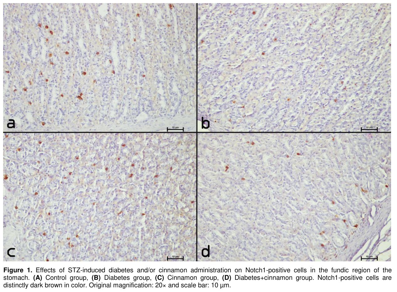
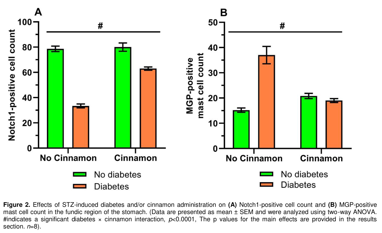
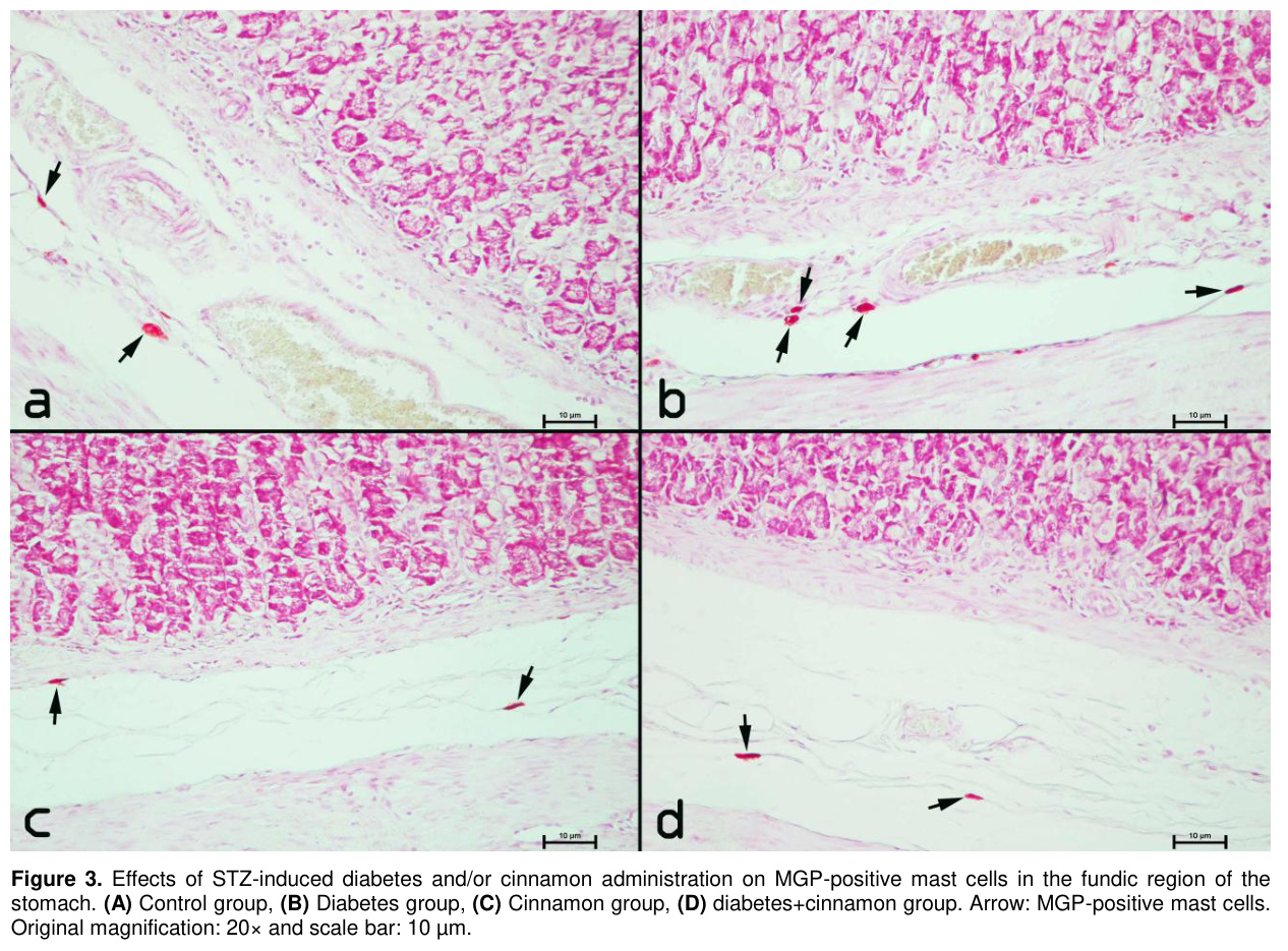
Notch1 Immunohistochemistry: Notch1 immunoreactivity was observed as distinct dark-brown DAB staining in scattered individual Notch1-positive cells throughout the gastric fundus (Figure 1). Regarding the number of Notch1-positive cells, significant main effects of diabetes (p<0.0001) and cinnamon (p<0.0001) were determined for Notch1-positive cell number. Because the diabetes × cinnamon interaction was also significant (p<0.0001, Figure 2A), simple-effects analyses were performed. Based on the estimated marginal means, the number of Notch1-positive cells was comparable between the cinnamon group (80.00±2.23) and the control group (78.60±2.23; p=0.660), indicating that cinnamon had no significant effect under non-diabetic conditions. In contrast, the number of Notch1-positive cells was significantly higher in the diabetes+cinnamon group (63.00±2.23) than in the diabetes group (33.40±2.23; p<0.0001), indicating a modulatory effect of cinnamon under diabetic conditions. The number of Notch1-positive cells was lower in the diabetes group (33.40±2.23) than in the control group (78.60±2.23; p<0.0001), indicating a marked diabetes-associated reduction in Notch1-positive cell number. The diabetes-induced decrease in Notch1-positive cell number was partially restored by cinnamon treatment; however, the diabetes+cinnamon group still had significantly fewer Notch1-positive cells than the cinnamon group (63.00±2.23 vs. 80.00±2.23; p<0.0001), suggesting that cinnamon partially ameliorated, but did not completely restore, the diabetes-associated reduction in Notch1-positive cell number.Mast Cell Histochemistry: In sections stained for MGP, MGP-positive mast cells with varying sizes and morphological features were observed. The cells were predominantly oval to round in shape, although spindle-shaped mast cells were also detected. All MGP-positive mast cells exhibited pinkish staining (Figure 3). These cells were predominantly localized in the lamina propria and submucosal regions of the gastric tissue and were frequently observed around blood vessels (Figure 3). With respect to the number of MGP-positive mast cells, significant main effects of diabetes (p<0.0001) and cinnamon (p=0.003) were observed. Because the diabetes × cinnamon interaction was also significant (p<0.0001, Figure 2B), simple-effects analyses were performed. Based on the estimated marginal means, the number of MGP-positive mast cells was significantly higher in the cinnamon group (20.80±1.88) than in the control group (15.20±1.88; p=0.044), indicating a stimulatory effect of cinnamon under non-diabetic conditions. In contrast, the number of MGP-positive mast cells was lower in the diabetes+cinnamon group (19.00±1.88) than in the diabetes group (37.00±1.88; p<0.0001), suggesting a modulatory effect of cinnamon under diabetic conditions. The diabetes group (37.00±1.88) had a significantly higher number of MGP-positive mast cells than the control group (15.20±1.88; p<0.0001), whereas no significant difference was observed between the diabetes+cinnamon group (19.00±1.88) and the cinnamon group (20.80±1.88; p=0.504), indicating that cinnamon counteracted the diabetes-associated increase in MGP-positive mast cells number.
Discussion
Notch signaling plays a pivotal role in cell fate and tissue-patterning decisions during development and continues to regulate tissue homeostasis and regeneration throughout life [31]. Growing evidence indicates that Notch signaling also contributes to gastric epithelial homeostasis through the regulation of gastric stem and progenitor cell proliferation and differentiation [32]. The Notch target gene Hes1 and Notch pathway components, including Notch1 and Jag2, have been identified in the developing gastric epithelium of embryonic mice [11]. In both human and mouse gastric tissues, Notch1 and Notch2 are the predominant Notch receptors, and their signaling has been implicated in regulating the proliferation of gastric antral and corpus progenitor cells [33]. Beyond gastrointestinal tissues, Notch signaling has also been implicated in metabolic regulation. Insulin secretion following postprandial glucose elevation is essential for maintaining glucose homeostasis by promoting glucose utilization and suppressing hepatic glucose production. In this context, Notch signaling has been implicated in hepatic glucose metabolism, with increased Notch activity associated with insulin resistance and enhanced hepatic glucose production [34]. Furthermore, inhibition of Notch signaling has been reported to improve insulin sensitivity in diet-induced insulin resistance, whereas Notch1 activation promotes insulin resistance [35]. Under diabetic conditions, Notch1 signaling has been reported to be altered in the retina. In STZ-induced diabetic mice, Notch1 expression was significantly reduced, accompanied by decreased DLL4 and p-Akt levels [36]. Similarly, suppression of Notch1 signaling in embryonic cardiac tissue has been reported in STZ-induced maternal diabetes, with reduced expression of Notch1 and downstream target genes, including Hey2, EfnB2, Nrg1, and Bmp10 [37]. Moreover, Yoon et al. [38] reported that high glucose increased Jagged1 expression and suppressed Notch1 signaling in endothelial cells. Zhao et al. [39] reported that STZ-induced diabetes increased miR-34a expression while reducing Notch1 expression in the vascular endothelium. Similarly, reduced Notch signaling activity in cardiac tissue has been reported in STZ-induced diabetic rats subjected to ischemia/reperfusion, accompanied by downregulation of Notch1 and other pathway components [40]. Consistent with these observations in other tissues, STZ-induced diabetes in the present study was associated with a marked reduction in the number of Notch1-positive cells in gastric tissue. Against this background, previous findings on the effects of cinnamon-derived compounds on Notch-related markers are particularly relevant. Previous studies have reported that cinnamaldehyde, a major bioactive component of cinnamon, can affect Notch-related signaling molecules, including Notch1, Jagged1, and NICD, in Schwann cells exposed to high glucose [41]. Interestingly, a botanical formulation containing Cinnamomum cassia has also been reported to modulate the Notch1/Hes5 signaling pathway, with medium- and high-dose treatment reversing the changes in Notch1 and Hes5 expression in the hippocampus [42]. Although these studies were conducted in different tissues and experimental settings, they suggest that cinnamon-related interventions may be associated with changes in Notch-related markers. Whether similar changes occur in diabetic gastric tissue remains unclear. In the present study, cinnamon treatment was associated with an increased number of Notch1-positive cells under diabetic conditions. Notably, cinnamon did not significantly alter Notch1-positive cell number under non-diabetic conditions, whereas it significantly increased Notch1-positive cell number under diabetic conditions. However, because Notch pathway activity was not directly assessed, these findings should be interpreted cautiously. These findings may reflect an association between cinnamon treatment and changes in Notch1-positive cell number but do not establish a direct effect on Notch signaling.Based on the staining characteristics of their cytoplasmic granules, mast cells exhibit histochemical heterogeneity and can be classified into three distinct subtypes according to Alcian blue–Safranin O staining: blue, red, and mixed. Blue mast cells contain Alcian blue-positive granules, red mast cells contain Safranin O-positive granules, whereas mixed mast cells exhibit both Alcian blue- and Safranin O-positive granules within the same cell [43]. These differential staining patterns reflect the histochemical heterogeneity of mast cell granules, which is associated with differences in their glycosaminoglycan composition and may vary according to tissue type and fixation conditions [44]. MGP staining is a classic histochemical technique used to differentially demonstrate DNA and RNA in tissue sections. In this staining method, methyl green selectively binds to nuclear DNA, producing a green coloration, whereas pyronin stains RNA-rich cytoplasmic components red or reddish-pink [45]. Due to their abundant cytoplasmic granules, mast cells can be readily identified morphologically using methyl green-pyronin (MGP) staining in paraffin-embedded tissue sections [46]. Although methyl green–pyronin (MGP) staining is not mast cell-specific, it has been used as an alternative histochemical method to demonstrate and morphologically evaluate mast cells in paraffin-embedded tissue sections [17]. In addition, in a study of rat lymph nodes, mast cells and their granules were morphologically identified using MGP staining, and mast cell numbers were evaluated [47]. Increased mast cell numbers have been reported in STZ-induced diabetic rats, particularly in the tongue, where a significant increase was observed after 4 weeks of diabetes [48]. In a rat model of type 2 diabetes, mast cell numbers were significantly increased in the islets of Langerhans compared with those in control animals [49]. Additionally, an increase in mast cell profiles was reported in the peripheral nerves of STZ-induced diabetic rats four weeks after the onset of diabetes, although this increase was attributed to changes in mast cell dimensions rather than proliferation [50]. Consistent with these reports, diabetes markedly increased MGP-positive mast cell number in animals not receiving cinnamon in the present study. Furthermore, cinnamon-derived type-A procyanidin polyphenols attenuated compound 48/80-induced mast cell degranulation and reduced histamine, β-hexosaminidase, and IL-4 levels in rat peritoneal mast cells [51]. Cinnamaldehyde, a major constituent of cinnamon, was also reported to dose-dependently inhibit antigen-induced degranulation and suppress extracellular Ca²⁺ influx in mucosal-type mast cells [52]. Although these studies evaluated mast cell activity and degranulation rather than mast cell number, they demonstrate that cinnamon-derived compounds can influence mast cell biology. In the present study, the effect of cinnamon on MGP-positive mast cell number differed according to diabetic status, with an increase under non-diabetic conditions and a significant reduction under diabetic conditions.
Notch signaling has been shown to enhance the antigen-presenting capacity of mast cells by inducing MHC-II and OX40L expression and promoting CD4⁺ T-cell activation and Th2 differentiation [53]. In addition, Notch signaling has been shown to enhance FcεRI-mediated cytokine production by mast cells through both direct and indirect mechanisms [54]. Honjo et al. (2017) reported that pharmacologic inhibition of Notch signaling suppressed food antigen-induced intestinal mast cell hyperplasia, supporting a role for Notch signaling in promoting mucosal mast cell differentiation and accumulation [55]. However, a previous study reported that Notch2 deficiency in bone marrow-derived cells led to an increase in intestinal mast cells, particularly in the lamina propria of the small intestine [56]. Furthermore, Notch and TGF-β signaling have been shown to act in a mutually dependent manner during the differentiation and maturation of mucosal mast cells. Notably, the concomitant activation of Notch and TGF-β signaling has been reported to promote the expression of characteristic mast cell markers, including mouse mast cell proteases (mMCP)-1 and mMCP-2, as well as αE integrin (CD103) [57]. In the present study, the decreased number of Notch1-positive cells and the increased number of MGP-positive mast cells observed in diabetic gastric tissue, together with the increase in Notch1-positive cells and reduction in MGP-positive mast cells following cinnamon treatment under diabetic conditions, may therefore be considered in the context of the reported relationship between Notch signaling and mast cell biology. Although these findings do not establish a direct relationship between Notch1-positive cells and MGP-positive mast cell numbers, the concurrent and opposite changes observed in these two cellular populations may suggest an association between changes in Notch1-positive cell number and MGP-positive mast cell number in gastric tissue.
Overall, STZ-induced diabetes was associated with a reduction in Notch1-positive cell number and an increase in MGP-positive mast cell number in gastric tissue. Cinnamon attenuated both diabetes-associated alterations, suggesting a diabetes-dependent modulatory effect on these cellular changes. The differing responses observed under diabetic and non-diabetic conditions further indicate that the gastric effects of cinnamon may depend on the underlying metabolic state. Further studies are needed to clarify the mechanisms underlying these effects.
References
- Reed J, Bain S, Kanamarlapudi V. A review of current trends with type 2 diabetes epidemiology, aetiology, pathogenesis, treatments and future perspectives. Diabetes Metab Syndr Obes 2021; 14: 3567-602.
- King GL, Loeken MR. Hyperglycemia-induced oxidative stress in diabetic complications. Histochem Cell Biol 2004; 122(4): 333-338.
- Brownlee M. The pathobiology of diabetic complications: A unifying mechanism. Diabetes 2004; 54: 1615-1625.
- Kornum DS, Krogh K, Keller J, et al. Diabetic gastroenteropathy: A pan-alimentary complication. Diabetologia 2025; 68(5): 905-919.
- Lee AA, Hasler WL. Diabetes and the stomach. Curr Treat Options Gastro 2017; 15(4): 441-459.
- Piyachaturawat P, Poprasit J, Glinsukon T, Wanichanon C. Gastric mucosal lesions in streptozotocin-diabetic rats. Cell Biol Int Rep 1988; 12(1): 53-63.
- Watanabe T, Asanuma A, Tanaka M, Akiba T, Koga T. Morphological study on the gastric mucosa in diabetes mellitus rats induced by streptozotocin. Exp Anim 1994; 43(5): 693-696.
- Kopan R. Notch signaling. Cold Spring Harb Perspect Biol 2012; 4(10): a011213-a011213.
- Kim W, Shin YK, Kim BJ, Egan JM. Notch signaling in pancreatic endocrine cell and diabetes. Biochem Biophys Res Commun 2010; 392(3): 247-251.
- Iso T, Kedes L, Hamamori Y. HES and HERP families: Multiple effectors of the notch signaling pathway. J Cell Physiol 2003; 194(3): 237-255.
- Kim TH, Shivdasani RA. Notch signaling in stomach epithelial stem cell homeostasis. J Exp Med 2011; 208(4): 677-688.
- Demitrack ES, Gifford GB, Keeley TM, et al. NOTCH1 and NOTCH2 regulate epithelial cell proliferation in mouse and human gastric corpus. Am J Physiol Gastrointest Liver Physiol 2017; 312(2): 133-144.
- Krystel-Whittemore M, Dileepan KN, Wood JG. Mast cell: A multi-functional master cell. Front Immunol 2016; 6: 620.
- Crivellato E, Ribatti D. The mast cell: An evolutionary perspective. Biol Rev 2010; 85(2): 347-360.
- Caughey GH. Mast cell proteases as pharmacological targets. Eur J Pharmacol 2016; 778: 44-55.
- Bischoff SC. Mast cells in gastrointestinal disorders. Eur J Pharmacol 2016; 778: 139-145.
- Genta RM, Turner KO, Collins MH, et al. Quantification of mucosal mast cells in the gastrointestinal tract: A primer for practicing pathologists. Arch Pathol Lab Med 2024; 148(2): e25-35.
- McKay DM, Bienenstock J. The interaction between mast cells and nerves in the gastrointestinal tract. Immunol Today 1994; 15(11): 533-538.
- Lin T, Befus A. Mast cells in mucosal Defensis and pathogenesis. In: Mestecky J, Lamm M, Strober W, et al. (Editors). Mucosal Immunity. Volume 1, United States of America: Elsevier Acedemic Press, 2002: 703-710.
- Setiyorini E, Qomaruddin MB, Wibisono S, et al. Complementary and alternative medicine for glycemic control of diabetes mellitus: A systematic review. J Public Health Res 2022; 11(3): 22799036221106582.
- Zhu C, Yan H, Zheng Y, et al. Impact of cinnamon supplementation on cardiometabolic biomarkers of inflammation and oxidative stress: A systematic review and meta-analysis of randomized controlled trials. Complement Ther Med 2020; 53: 102517.
- Mendis Abeysekera WPK, Arachchige SPG, Abeysekera WKSM, Ratnasooriya WD, Indeewari Medawatta HMU. Antioxidant and glycemic regulatory properties potential of different maturity stages of leaf of ceylon cinnamon (Cinnamomum zeylanicum blume) in vitro. Evid Based Complement Alternat Med 2019; 2019: 1-10.
- Lee MJ, Seo HJ, Hwang GS, et al. Molecular mechanism of Cinnamomum cassia against gastric damage and identification of active compounds. Biomolecules 2022; 12(4): 525.
- Han YM, Song MY, Lee DY, et al. Gastric Mucosal protective effects of Cinnamomum cassia in a rat model of ethanol-induced gastric injury. Nutrients 2023; 16(1): 55.
- Loganathan R, Novikova L, Boulatnikov IG, Smirnova IV. Exercise-induced cardiac performance in autoimmune (Type 1) diabetes is associated with a decrease in myocardial diacylglycerol. J Appl Physiol 2012; 113(5): 817-826.
- Hemayatkhah JV, Baqerzadeh P. The effect of cinnamon extract on ovary in adult rats treated lead acetate. J Anim Biol 2017; 9: 19.
- Çetin A, Vardı N, Orman D. Deneysel diyabetin sıçan kalp dokusunda meydana getirdiği histolojik değişiklikler üzerine aminoguanidinin iyileştirici etkileri. İnönü Üniv Sağlık Hiz MYO Derg 2013; 1(1): 17-26.
- True L. Principles of immunohistochemistry. In: True L, (Editor). Atlas of Diagnostic Immunohistopathology. New York: Gower Medical Publishing; 1990: 16-22.
- Bancroft JD, Cook HC. Manual of Histological Techniques. New York: Churchill Livingstone, 1984.
- Ülker Ertuğrul N, Gökdere E, Keskin F, et al. Comparison of meteorin-like protein infusion vs. moderate-intensity exercise on cardiac mast cell and plasma cell dynamics and musclin levels in female rats. Turk Kardiyol Dern Ars 2025; 53(6): 381-387.
- Bray SJ, Bigas A. Modes of Notch signalling in development and disease. Nat Rev Mol Cell Biol 2025; 26(7): 522-537.
- Demitrack ES, Samuelson LC. Notch regulation of gastrointestinal stem cells. J Physiol 2016; 594(17): 4791-4803.
- Demitrack ES, Samuelson LC. Notch as a driver of gastric epithelial cell proliferation. Cell Mol Gastroenterol Hepatol 2017; 3(3): 323-330.
- Pajvani UB, Qiang L, Kangsamaksin T, et al. Inhibition of notch uncouples akt activation from hepatic lipid accumulation by decreasing mTorc1 stability. Nat Med 2013; 19(8): 1054-1060.
- Pajvani UB, Shawber CJ, Samuel VT, et al. Inhibition of Notch signaling ameliorates insulin resistance in a FoxO1-dependent manner. Nat Med 2011; 17(8): 961-967.
- Qin X, Zhang Z, Xu H, Wu Y. Notch signaling protects retina from nuclear factor-κB- and poly-ADP-ribose-polymerase-mediated apoptosis under high-glucose stimulation. Acta Biochim Biophys Sin 2011; 43(9): 703-711.
- Basu M, Zhu JY, LaHaye S, et al. Epigenetic mechanisms underlying maternal diabetes-associated risk of congenital heart disease. JCI Insight 2017; 2(20): e95085.
- Yoon C, Choi Y, Kim H. High glucose condition induced jagged–1 overexpression and notch inhibition, resulting in the aberrant endothelial tube formation: A novel mechanism of diabetic microvasculopathy in a model of human angiogenesis. J Am Coll Cardiol 2013; 61(10): E2086.
- Zhao D, Wang NS, Chen F, et al. Intravenous injection of mir-34a inhibitor alleviates diabetes mellitus-induced vascular endothelial dysfunction by targeting NOTCH1. Exp Clin Endocrinol Diabetes 2019; 127(04): 255-262.
- Wu F, Yu B, Zhang X, Zhang Y. Cardioprotective effect of Notch signaling on the development of myocardial infarction complicated by diabetes mellitus. Exp Ther Med 2017; 14(4): 3447-3454.
- Liu D, Liang XC, Sun Y, Wu YN, Zhang H. Combination of quercetin, hirudin and cinnamaldehyde promotes schwann cell differentiation and myelination against high glucose by inhibiting ERK signaling pathway. Chin J Integr Med 2020; 26(8): 591-598.
- Liu B, Zhang Q, Ke C, et al. Ginseng-angelica-sansheng-pulvis boosts neurogenesis against focal cerebral ischemia-induced neurological deficiency. Front Neurosci 2019; 13: 515.
- El Sayed SO, Dyson M. Histochemical heterogeneity of mast cells in rat dermis. Biotech Histochem 1993; (68): 326-32.
- West PW, Bulfone-Paus S. Mast cell tissue heterogeneity and specificity of immune cell recruitment. Front Immunol 2022; 13: 932090.
- Luna LG. Histopathologic Methods and Color Atlas of Special Stains and Tissue Artifacts, Maryland: American Histolabs, 1992.
- Chen CM, Chang HL. Methyl green-pyronin as a stain for mast cells in paraffin sections. Stain Technology 1963; 38: 133-135.
- Dabak DO, Aydin G, Ozguner M. Dynamics of mast cells in lymph node following antigenic stimulation. Anat Histol Embryol 2004; 33(1): 5-10.
- Batbayar B, Somogyi J, Zelles T, Fehér E. Immunohistochemical analysis of substance P containing nerve fibres and their contacts with mast cells in the diabetic rat’s tongue. Acta Biol Hung 2003; 54(3-4): 275-284.
- Abdel-Hamid AAM, Firgany AEDL. Increased mast cell number is associated with a decrease in beta-cell mass and regeneration in type 2 diabetic rats. Acta Histochem 2019; 121(4): 508-515.
- Jakobsen J, Jensen HD, Sidenius P. Change in mast cell dimensions increases the profile number in sections of peripheral nerve of diabetic rats. Acta Neuropathol 1982; 56(2): 157-160.
- Kandhare AD, Aswar UM, Mohan V, Thakurdesai PA. Ameliorative effects of type-A procyanidins polyphenols from cinnamon bark in compound 48/80-induced mast cell degranulation. Anat Cell Biol 2017; 50(4): 275.
- Kageyama-Yahara N, Wang X, Katagiri T, et al. Suppression of phospholipase Cγ1 phosphorylation by cinnamaldehyde inhibits antigen-induced extracellular calcium influx and degranulation in mucosal mast cells. Biochem Biophys Res Commun 2011; 416(3-4): 283-288.
- Nakano N, Nishiyama C, Yagita H, et al. Notch signaling confers antigen-presenting cell functions on mast cells. J Allergy Clin Immunol 2009; 123(1): 74-81.e1.
- Nakano N, Nishiyama C, Yagita H, et al. Notch signaling enhances FcεRI-mediated cytokine production by mast cells through direct and indirect mechanisms. J Immunol 2015; 194(9): 4535-4544.
- Honjo A, Nakano N, Yamazaki S, et al. Pharmacologic inhibition of Notch signaling suppresses food antigen-induced mucosal mast cell hyperplasia. J Allergy Clin Immunol 2017; 139(3): 987-996.
- Sakata-Yanagimoto M, Sakai T, Miyake Y, et al. Notch2 signaling is required for proper mast cell distribution and mucosal immunity in the intestine. Blood 2011; 117(1): 128-134.
- Nakano N, Saida K, Hara M, et al. Mucosal Mast Cell–specific gene expression is promoted by interdependent action of notch and TGF-β signaling. J Immunol 2021; 207(12): 3098-3106.
- Effects of Warfarin, Aspirin and Sulfanilamide on Protrombin Time in Healthy Mice
- SIĞIRLARIN BAZI ÖNMİDE HASTALIKLARI VE PRİMER KETOZİSİN KARACİĞER İŞLEVLERİ ÜZERİNE ETKİSİ
- ELAZIĞ YÖRESİ KOYUNLARINDA MİDE-BAĞIRSAK NEMATODLARININ YAYGINLIĞI *
- ELAZIĞ YÖRESİ SIĞIRLARINDA SİNDİRİM SİSTEMİ NEMATODLARININ YAYGINLIĞI *
- The Effects of Furosemide and Indomethacin on the Serum Osmolality and Electrolyte Levels in the Sheep