
Evcil Güvercinlerde (Columba livia domestica) Avian Pox Virüsü Enfeksiyonu: Histopatolojik ve Moleküler Bulgularla Bir Olgu Sunumu
Volume 40, Issue 3
October 2026 · Pages 297-300
Evcil Güvercinlerde (Columba livia domestica) Avian Pox Virüsü Enfeksiyonu: Histopatolojik ve Moleküler Bulgularla Bir Olgu Sunumu
Cutaneous Avian Poxvirus Infection in a Domestic Pigeon (Columba livia domestica): A Case Report with Histopathological and Molecular Findings
Canan AKDENİZ İNCİLİ1, Yesari ERÖKSÜZ1, Hakan IŞIDAN2
2Cumhuriyet University, Faculty of Veterinary Medicine, Department of Virology, Sivas, TÜRKİYE
On beş–yirmi günlük erkek evcil bir güvercinde (Columba livia domestica) deri avian poksvirüs enfeksiyonu tanımlandı. Klinik muayenede, baş ve ayaklarda 0,2-2 mm çapında sert, sarı, proliferatif nodüller saptandı. Lezyonlu dokular kriyostat ve parafin kesitler histopatolojik olarak incelendi. Mikroskobik değerlendirmede, epidermis ve foliküler epitelde belirgin eozinofilik intrasitoplazmik inklüzyon cisimleri (Bollinger cisimleri) ile birlikte belirgin hidropik dejenerasyon ve epitel hiperplazisi saptandı. Etiyolojik doğrulama için, avian poksvirüs DNA'sını hedef alan polimeraz zincir reaksiyonu (PCR) yapıldı ve pozitif sonuç alındı. Karakteristik makroskobik bulgular, destekleyici histopatoloji ve moleküler doğrulama bir araya getirildiğinde, kutanöz avian pox tanısı konuldu. Bu vaka, evcil güvercinlerde avian poksvirüs enfeksiyonunun doğru tanısı için klinik bulgular, histopatoloji ve PCR'nin entegre edilmesinin önemini vurgulamaktadır.
A cutaneous avian poxvirus infection was diagnosed in a 15–20-day-old male domestic pigeon (Columba livia domestica). Clinical examination revealed firm, yellow, proliferative nodules measuring 0.2–2 mm in diameter on the head and feet. Lesion tissues were processed by frozen sectioning and routine paraffin histopathology. Microscopic evaluation showed marked hydropic degeneration and epithelial hyperplasia, with distinct eosinophilic intracytoplasmic inclusion bodies (Bollinger bodies) in the epidermis and follicular epithelium. For etiological confirmation, polymerase chain reaction (PCR) targeting avian poxvirus DNA was performed and revealed a positive result. Taken together, the characteristic gross presentation, supportive histopathology, and molecular confirmation established the diagnosis of cutaneous avian pox. This case emphasizes the value of integrating clinical findings, histopathology, and PCR for accurate diagnosis of avian poxvirus infection in domestic pigeons.
Introduction
Avian pox is a slow-progressing, contagious viral disease caused by Avian poxvirus, a double-stranded DNA virus within the family Poxviridae, and it affects a broad range of avian hosts, including the domestic pigeon (Columba livia domestica) [1]. In pigeons, infection most commonly presents as cutaneous, proliferative nodules that develop on unfeathered skin, particularly around the eyelids, beak commissures, and legs. Although the cutaneous form is often described as self-limiting, the disruption of the epidermal barrier and local tissue damage may facilitate secondary bacterial colonization, which can increase morbidity especially in juveniles or otherwise immunocompromised birds.Diagnosis of avian pox relies heavily on pathology, because the disease produces a highly recognizable histologic pattern. Histopathological examination typically reveals marked epidermal hyperplasia, ballooning (hydropic) degeneration, and eosinophilic intracytoplasmic inclusion bodies (Bollinger bodies) within affected epithelial cells [2,3]. When inclusions are well developed, these changes are considered pathognomonic and support confident differentiation from other proliferative or ulcerative dermatoses. However, lesion stage and concurrent secondary infection can occasionally obscure classic features, making molecular confirmation an important complement to histology. In this setting, PCR targeting the conserved P4b gene is widely used for sensitive detection and for supporting strain-level differentiation among avian poxviruses, including fowlpox- and avian pox-associated viruses [4].
Beyond individual case diagnosis, avian poxvirus infections remain relevant from a surveillance perspective. Recent molecular investigations from Basrah, Iraq, for example, have demonstrated ongoing circulation of avian pox virus in backyard/free-ranging pigeon populations using sequence-based confirmation [5]. Such reports reinforce the value of continuous monitoring across domestic and wild bird interfaces, where management practices, vector exposure, and mixed-species contact may influence virus maintenance and spread.
Against this background, the present report describes cutaneous avian pox in a juvenile domestic pigeon, supported by gross lesions, histopathological hallmarks, and PCR-based detection. The case contributes to the documentation of avian poxvirus infection in non-commercial avian hosts and underscores the practical diagnostic advantage of integrating pathology and molecular methods in veterinary virology.
Case Presentation
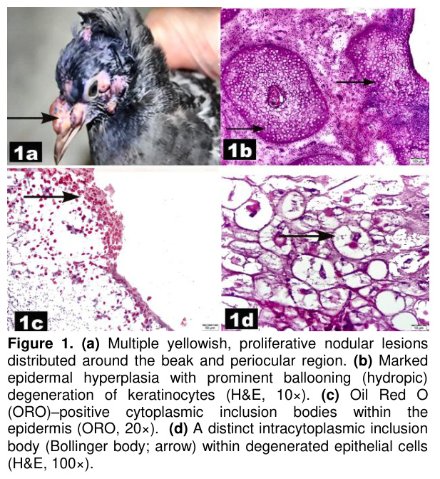
A 15–20-day-old male domestic pigeon (Columba livia domestica) was submitted alive to the Department of Pathology, Faculty of Veterinary Medicine, Fırat University. Prior to necropsy, the pigeon was humanely euthanized by cervical dislocation. During the systemic necropsy examination, numerous yellowish, firm, proliferative nodules ranging in diameter from 0.2 to 2 mm were observed; these were predominantly found in the head region particularly around the beak and also on the feet (Figure 1a). Apart from the liver, which appeared generally pale, no lesions were observed in the other tissues.Tissue samples collected during necropsy were fixed in 10% neutral-buffered formalin, routinely processed, and embedded in paraffin. Sections were cut at a thickness of 4–5 µm and stained with hematoxylin and eosin (H&E) for light microscopic examination.
For lipid histochemistry, additional samples from the cutaneous nodules were processed as frozen sections. Briefly, the tissues were embedded in optimal cutting temperature (OCT) compound, cryosectioned at a thickness of 6–8 µm, and air-dried. The sections were subsequently stained with Oil Red O (ORO) according to a standard protocol.
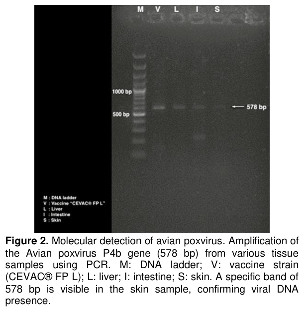
DNA Extraction: DNA extraction from the live vaccine strain (CEVAC® FP L) and the tissue samples (liver, intestine, and skin) was performed using the High Pure PCR Template Preparation Kit (Roche), according to the manufacturer's recommendation.
PCR: The obtained DNA samples were amplified using the FPV F (5'-CAGCAGGTGCTAAACAACAA-3') and FPV R (5'-CGGTAGCTTAACGCCGAATA-3') primers specific to the P4b gene [6]. The PCR reaction for disease diagnosis was carried out in a total of 50 µL PCR mix, consisting of 5 µL DNA, 5 µL 10xPCR Buffer (100 mM Tris-HCl, pH: 8.0, 500 mM potassium chloride, 15 mM magnesium chloride), 2 mM of each of the 4 deoxynucleotides, 1U Taq DNA polymerase (Fermentas), and 20 pM forward and reverse primers. Amplification was performed with 35 cycles at 94°C for 3 minutes, 95°C for 1 minute, 55°C for 2 minutes, 72°C for 1 minute, and a final extension at 72°C for 10 minutes. 10 µL of PCR product was run on a 2% agarose gel and visualized with ethidium bromide staining under UV light.
Histopathological Findings: The epidermis showed severe, diffuse hydropic degeneration, accompanied by marked epithelial hyperplasia. Numerous eosinophilic intracytoplasmic inclusion bodies (Bollinger bodies) were observed within the cytoplasm of keratinocytes (Figures 1b–d).
ORO staining demonstrated lipid positivity within the intracytoplasmic inclusion bodies (Bollinger bodies) of degenerated epithelial cells.
Molecular Findings: In the examined tissues, the expected 578-bp P4b amplicon was detected in the skin sample, supporting the cutaneous localization of infection in this case (Figure 2).
Discussion
This case report documents a cutaneous avian poxvirus infection (pigeonpox) in a juvenile domestic pigeon, confirmed through gross pathology, histopathology, and PCR. Juvenile birds are particularly susceptible to avian poxvirus infections because of immature immune function and limited maternal antibody protection [1,7]. In young pigeons, the relatively thin and incompletely keratinized integument may further facilitate viral entry and local replication, which fits well with the typical predilection of avian poxviruses for keratinized epithelium and exposed body sites. In the present case, the distribution of lesions on the head and feet is therefore consistent with established clinicopathological patterns of cutaneous avian pox in pigeons [1].Grossly, the firm, proliferative nodules reflect the virus’s capacity to induce epithelial proliferation at sites that are vulnerable to microtrauma and arthropod contact. Histopathologically, the lesions were characterized by pronounced epidermal and follicular hyperplasia accompanied by marked hydropic degeneration, together with eosinophilic intracytoplasmic inclusion bodies (Bollinger bodies). The identification of these inclusions remains one of the most informative morphologic hallmarks of avian poxvirus infection and is widely regarded as pathognomonic when present [2,3]. The degree of epithelial hyperplasia observed in this case is biologically plausible in light of poxvirus pathobiology: avian poxviruses, like other poxviruses, can encode EGF-like proteins that stimulate mitogenic signaling in keratinocytes, supporting a cellular environment favorable for viral replication [8]. Additionally, the ORO positivity of inclusion bodies in this case is in agreement with their lipid-rich viroplasmic composition, a feature that has been described in histochemical and ultrastructural work on poxvirus replication “factories” [2,8].
Although the classic histologic constellation is highly suggestive, practical diagnostic experience shows that lesions encountered at an early stage, in regression, or complicated by secondary bacterial colonization may not always display abundant inclusions or may show overlapping inflammatory and proliferative features. For this reason, molecular confirmation can be particularly valuable when morphology alone is equivocal. In the present case, PCR amplification of the conserved P4b gene confirmed the presence of Avian poxvirus DNA, underscoring PCR as a sensitive and specific method for confirmation and surveillance [4].
From an epidemiological standpoint, recent surveillance such as studies reported from Basrah, Iraq suggests that avian pox continues to circulate in backyard pigeon populations and that circulating strains may exhibit notable genetic diversity [5]. Such findings support the broader relevance of continued monitoring and genotyping, both for improving local disease control and for informing regionally appropriate prevention strategies. While avian poxviruses are generally considered host-adapted, reports of high sequence similarity between some avian pox and fowl pox strains (in some comparisons exceeding 95% homology) raise the possibility that cross-species transmission could occur under certain ecological or management conditions, particularly when multiple avian hosts are maintained in close proximity [1,5]. Experimental work also supports this potential: pigeon-derived avian poxvirus has been shown to infect chickens and Japanese quail, producing comparable cutaneous lesions and histopathologic changes [9]. In natural settings, mosquitoes and other hematophagous insects are central to mechanical transmission between hosts, especially in open-loft systems and mixed backyard aviaries, and fomite-based spread via contaminated surfaces, feeders, or handling likely contributes to horizontal dissemination. Comparable pox-like cutaneous lesions have also been described in free-ranging juvenile pigeons, reinforcing that young birds may present with extensive proliferative nodules even outside commercial settings [10]. Moreover, cases showing both cutaneous (dry) and diphtheritic (wet) forms in pigeons have been reported, underlining the clinical importance of monitoring for mucosal involvement even when initial lesions appear restricted to the skin [11].
Clinically, the apparent lack of mucosal involvement in this pigeon likely limited disease severity. This contrasts with the diphtheritic form, where oropharyngeal and upper respiratory involvement can lead to substantial morbidity and increased mortality due to impaired respiration and feeding [12]. In this context, the restricted distribution of lesions in the current case may reflect early recognition, a comparatively lower virulence phenotype of the infecting strain, or host–environment interactions that limited progression.
Effective prevention in pigeon flocks is rarely achieved through a single measure; instead, it generally requires a layered approach combining vector control (mosquito reduction and elimination of breeding sites), strict hygiene and biosecurity to reduce fomite transmission, and quarantine of newly introduced birds. Where vaccination is feasible, wing-web inoculation is commonly used in pigeons and may be most effective when applied prior to periods of high vector activity, integrated with management practices that reduce crowding and contact opportunities.
Taken together, this case highlights how a careful combination of clinical observation, histopathologic confirmation (including Bollinger bodies), and PCR-based detection can provide a robust diagnosis of cutaneous avian pox in juvenile pigeons. Beyond documenting an individual animal’s disease, such integrated diagnosis also supports practical flock-level decision-making—helping clinicians and owners recognize the infection early, implement appropriate control measures, and reduce the likelihood of spread in backyard or multi-species settings where the consequences of delayed recognition can be disproportionately high.
References
- Marlier D, Vindevogel H. Viral infections in pigeons. Vet J 2006; 172(1): 40-51.
- Hibl BM, Blackwood RS, Simons BW, Collins DE. Poxvirus Infection in a Colony of Laboratory Pigeons (Columba livia). Comp Med 2019; 69(3): 179-183.
- Shalaby SM, Awadin WF, Hamed MF, et al. Pathological and ultrastructural characteristics of Newcastle and pox diseases in naturally infected pigeons in Egypt. Adv Anim Vet Sci 2021; 9(11): 1995-2004.
- Mohamed RI, Elsamadony HA, Alghamdi RA, et al. Molecular and pathological screening of the current circulation of fowlpox and pigeon pox virus in backyard birds. Poult Sci 2024; 103:104249.
- Khaleefah IA, Al-Tameemi HM, Kraidi QA, et al. Clinical and molecular detection of fowl pox in domestic pigeons in Basrah Southern of Iraq. Korean J Vet Res 2024; 64(1): e7.
- Lee LH, Lee KH. Application of the polymerase chain reaction for the diagnosis of fowl poxvirus infection. J Virol Methods 1997; 63(1-2): 113-119.
- Tripathy DN, Reed WM. Pox. In: Saif YM, Barnes HJ, Glisson JR, et al (Editors). Diseases of Poultry. 1st Edition, Ames Iowa: Iowa State University Press; 2003: 253-269.
- McFadden G. Poxvirus tropism. Nat Rev Microbiol 2005; 3(3): 201-213.
- Siddique MAB, Hossain MA. Cross-infection of pigeonpox virus in Japanese quail and chicken under experimental conditions. Int J Poult Sci 2011; 10(3): 207-211.
- Bwala DG, Fasina FO, Duncan NM. Avian poxvirus in a free-range juvenile speckled (rock) pigeon (Columba guinea). J S Afr Vet Assoc 2015; 86(1): 1-4.
- Abbiw RK, Amoako KK, Enyetornye B, et al. Isolation and molecular detection of pigeonpox virus in a pigeon with both cutaneous and diphtheritic forms of pigeon pox disease in Ghana. Vet Med Int 2025; 7523480: 7.
- Ramesh S, Soundararajan C, Selvaraj J, Subapriya S. Avian pox in a blue rock pigeon – a case report. Int J Curr Microbiol Appl Sci 2018; 7(12): 3690-3692.