
Farelerde Sisplatine Bağlı Nörotoksisiteye Karşı Safranalın Antioksidan ve Antiinflamatuvar Mekanizmalar Yoluyla Koruyucu Etkileri
Cilt 40, Sayı 3
Ekim 2026 · Sayfalar 233-241
Farelerde Sisplatine Bağlı Nörotoksisiteye Karşı Safranalın Antioksidan ve Antiinflamatuvar Mekanizmalar Yoluyla Koruyucu Etkileri
Protective Effects of Safranal Against Cisplatin-Induced Neurotoxicity through Antioxidant and Anti-inflammatory Mechanisms in Mice
Sedat GÖKMEN1, Mehmet Sait ATAR2, İrfan ÇINAR3, Muhammed ETYEMEZ4, Musa TATAR5
2Kastamonu University, Faculty of Veterinary Medicine, Department of Histology and Embryology, Kastamonu, TÜRKİYE
3Kastamonu University, Faculty of Medicine, Department of Medical Pharmacology, Kastamonu, TÜRKİYE
4Kastamonu University, Faculty of Veterinary Medicine, Department of Physiology, Kastamonu, TÜRKİYE
5Karabük University, Faculty of Medicine, Department of Histology and Embryology, Karabük, TÜRKİYE
Sisplatin (CIS), testis, over, mesane ve akciğer kanserleri dâhil olmak üzere çeşitli solid tümörlerin tedavisinde kullanılan platin bazlı bir kemoterapötik ilaçtır. Ancak klinik kullanımı, özellikle nörotoksisite olmak üzere doza bağlı yan etkiler nedeniyle sınırlanmaktadır. Bu çalışmada, farelerde safranalın sisplatin kaynaklı nörotoksisiteye karşı koruyucu etkileri araştırılmıştır. Erkek Balb/c fareler rastgele altı gruba ayrıldı (her grupta n = 7): kontrol, taşıt (vehicle) kontrol, CIS, CIS + safranal (0.1 mL/kg), CIS + safranal (0.2 mL/kg) ve CIS + safranal (0.4 mL/kg). Safranal, 14 gün boyunca günde bir kez intraperitoneal olarak uygulandı. Nörotoksisite oluşturmak amacıyla, 11. günde CIS ve CIS + safranal gruplarına tek doz intraperitoneal CIS (7 mg/kg) uygulandı. Oksidatif stres parametreleri olan malondialdehit (MDA) düzeyi, süperoksit dismutaz (SOD) aktivitesi ve glutatyon (GSH) aktivitesi ELISA kitleri kullanılarak ölçüldü. NF-κB, TNF-α, IL-1β ve IL-6 mRNA ekspresyon düzeyleri RT-qPCR yöntemiyle analiz edilmiştir. Histopatolojik bulgular, hematoksilen-eozin ile boyanmış beyin kesitlerinde yarı kantitatif skorlama yöntemi kullanılarak değerlendirildi. Sisplatin uygulaması, beyin dokusunda MDA düzeylerini artırırken, SOD ve GSH aktivitelerini azalttı (p<0,05) ve NF-κB, TNF-α, IL-1β ve IL-6 mRNA ekspresyon düzeylerini artırdı (p<0.05). Histopatolojik incelemede, safranal uygulanan gruplarda CIS grubuna kıyasla doku hasarının azaldığı gözlendi. Sonuç olarak, safranal oksidatif stres ve inflamasyonu azaltarak sisplatine bağlı nörotoksisiteye karşı umut verici nöroprotektif etkiler göstermiştir. Ancak, bu etkinin doğrulanması ve etki mekanizmalarının daha ayrıntılı olarak ortaya konulabilmesi için farklı dozlar, uygulama süreleri ve deneysel modellerde ileri preklinik çalışmaların yapılması gerekmektedir.
Cisplatin (CIS) is a platinum-based chemotherapeutic agent used to treat various solid tumors, including testicular, ovarian, bladder, and lung cancers. However, its clinical application is limited by dose-dependent adverse effects, particularly neurotoxicity. This study investigated safranal's protective effects against CIS-induced neurotoxicity in mice. Male Balb/c mice were randomly assigned to six groups (n = 7 per group): control, vehicle control, CIS, CIS + safranal (0.1 mL/kg), CIS + safranal (0.2 mL/kg), and CIS + safranal (0.4 mL/kg). Safranal was administered intraperitoneally once daily for 14 days. On day 11, the CIS and CIS + safranal groups received a single intraperitoneal injection of CIS (7 mg/kg) to induce neurotoxicity. Oxidative stress parameters, including malondialdehyde (MDA) levels, superoxide dismutase (SOD) activity, and glutathione (GSH) activity, were measured using ELISA kits. NF-κB, TNF-α, IL-1β, and IL-6 mRNA expression levels were analyzed using RT-qPCR. Histopathological alterations were evaluated in hematoxylin and eosin-stained brain sections using a semi-quantitative scoring system. CIS exposure increased MDA levels and decreased SOD and GSH activities in brain tissue (p<0.05) and increased mRNA expression of NF-κB, TNF-α, IL-1β, and IL-6 (p<0.05). Histopathological examination showed reduced tissue damage in safranal treatment groups. In conclusion, safranal demonstrated neuroprotective effects against CIS-induced neurotoxicity by reducing oxidative stress and inflammation. However, further preclinical studies employing different doses, treatment durations, and experimental models are warranted to confirm these findings and elucidate the underlying molecular mechanisms.
Giriş
Chemotherapy is a widely used and effective approach for cancer treatment. However, these drugs also induce adverse effects in healthy tissue [1]. Cisplatin (Cl2H6N2Pt) (CIS) , a platinum-based chemotherapeutic drug, is a widely used treatment for various malignancies, including lung, ovarian, head and neck, testicular, and non-small-cell lung cancers. However, despite its substantial clinical success, its therapeutic application is considerably restricted by dose-dependent adverse effects, including neurotoxicity, ototoxicity, nephrotoxicity, and testicular toxicity. These toxicities not only compromise the quality of life of patients but also frequently necessitate dose reduction or discontinuation of therapy, thereby limiting its overall efficacy [2,3]. Neurotoxicity is one of the most significant complications. Clinical evidence indicates that approximately 40–50% of patient receiving cisplatin therapy develop chemotherapy-induced peripheral neuropathy [1]. Among these patients, painful peripheral neuropathy has been reported with a prevalence of up to 85%, depending on the patient population, cumulative cisplatin dose, and assessment criteria [2,3]. These symptoms may emerge several years after cisplatin treatment, significantly impacting cancer survivors’ quality of life [1-3]. The underlying mechanisms of neurotoxicity caused by cisplatin are multifactorial and involve oxidative stress, inflammation, and apoptosis. At the cellular level, toxicity primarily arises from alterations in transcription and DNA replication. Furthermore, accumulating evidence suggests that oxidative stress and inflammation are the major contributors to CIS-induced toxicity, with the predominant mechanism of damage involving excessive generation of reactive oxygen species, depletion of endogenous antioxidant defenses, and increased levels of pro-inflammatory cytokines [1]. Previous studies have reported that CIS causes oxidative stress and inflammation in brain tissue, leading to nerve damage [4-6]. As there are currently no Food and Drug Administration-approved therapies for CIS-induced neurotoxicity, the development of alternative approaches capable of modulating the underlying mechanisms is critical [2,3]. In the literature, various antioxidants and anti-inflammatory supporting substances have been investigated to reduce the undesirable toxic effects of cisplatin, a chemotherapeutic agent widely used in cancer treatment.One of the objectives of preclinical studies in this area is to identify a treatment method that offers sufficient protection against CIS-induced neurotoxicity. Consequently, employing an agent with antioxidant, anti-inflammatory, and anti-cancer properties as a protective measure would be promising in countering the neurotoxicity of CIS [1]. Extensive research has focused on utilizing plants as medicinal sources for many diseases, aiming to mitigate the adverse effects associated with chemical medications [7]. In recent years, increasing attention has been directed toward plant-derived bioactive compounds because of their diverse pharmacological activities, including antioxidant, anti-inflammatory, and anti-apoptotic effects. These compounds have been widely investigated as potential adjunctive agents to alleviate the toxic side effects associated with chemotherapeutic drugs, particularly cisplatin-induced organ toxicity [8].
Safranal, derived from the stigmas of Crocus sativus L., possesses potential therapeutic properties [9,10]. Safranal is effectively used in different areas, such as pharmaceuticals and nutraceuticals [11,12]. Safranal (C10H14O, 2,6,6-trimethyl-1,3-cyclohexadiene-1-carboxaldehyde) [13], a principal component of saffron, is a potent monoterpene aldehyde exhibiting a wide range of pharmacological properties, including anti-inflammatory, antioxidant, anti-apoptotic and antidepressant effects. Numerous studies have demonstrated the neuroprotective potential of safranal [14-16]. Safranal is specifically recognized for its neuroprotective activity against neurological disorders. To date, published findings suggest its potential efficacy in mitigating conditions such as epilepsy, stroke, multiple sclerosis, Parkinson’s disease and Alzheimer’s disease [7,10].
However, the protective role of safranal against cisplatin-induced neurotoxicity has not been sufficiently clarified using combined oxidative stress, inflammatory gene expression, and histopathological analyses. Therefore, this study aimed to investigate the neuroprotective effects of safranal against CIS-induced neurotoxicity in mice by evaluating oxidative stress parameters, inflammatory signaling pathways, and histopathological alterations in brain tissue.
Gereç ve Yöntem
Research and Publication Ethics: The Local Ethics Committee for Animal Experiments approved this study at Kastamonu University (Approval number: 2026/01/04).Chemicals: Safranal (CAS No: 116-26-7, purity ≥98%, density = 0.98 g/mL; molecular weight = 150.22 g/mol) and cisplatin (CAS No: 15663-27-1, purity ≥99%, molecular weight = 300.05 g/mol) were obtained from Sigma Aldrich Chemical Co.
Experimental Animals: In this study, we used 42 male Balb/c mice (6–8 weeks old, weighing 25–30 g) obtained from the Laboratory Animals and Research Laboratory, Ankara University, Türkiye. The animals were fed under normal temperature conditions (21–24°C), 40–45% humidity, and light-controlled (12 h light/12 h dark) conditions in separate groups before the experiments at the Kastamonu University Experimental Animals Research and Application Center, Kastamonu, Türkiye. The animals were maintained on a standard pellet diet and given water ad libitum during the experimental period. To minimize the influence of circadian variations, safranal was administered daily, and the single CIS injection (on day 11) was administered between 09:00 and 11:00 a.m. throughout the experimental period.
Experimental Design and Groups: Following a seven-day acclimation period, the mice were randomly allocated into six groups (n = 7) as detailed below:
Control (C): Animals received 0.2 mL of FTS intraperitoneally (i.p.) for 14 days.
Vehicle Control (VC): Animals received 0.2 mL of liquid paraffin (solvent for safranal) i.p. for 14 days [17].
Cisplatin (CIS): Animals received a single i.p. dose of cisplatin (7 mg/kg) on day 11 [18].
Cisplatin + Safranal 0.1 mL/kg (CIS+S 0.1): Animals were administered safranal i.p. at a dose of 0.1 mL/kg for 14 days [19], and CIS (7 mg/kg, i. p.) was administered only on day 11.
Cisplatin + Safranal 0.2 mL/kg (CIS+S 0.2): Animals were administered safranal at a dose of 0.2 mL/kg (i. p.) for 14 days [19], and CIS (7 mg/kg, i. p.) was administered only on day 11.
Cisplatin + Safranal 0.4 mL/kg (CIS+S 0.4): Animals were administered safranal at a dose of 0.4 mL/kg (i. p.) for 14 days [19], and CIS (7 mg/kg, i. p.) was administered only on day 11.
On the 15th day of treatment, the animals were euthanized by cervical dislocation under high-dose general anesthesia with xylazine (10 mg/kg, i.p.) and ketamine (200 mg/kg, i.p.). Immediately after euthanasia, the whole brain was carefully removed, and the brain tissue was collected. A portion of the samples was preserved at −80°C until oxidative stress and RT-qPCR analyses, whereas the remaining tissue samples were fixed in 10% neutral-buffered formalin for histopathological evaluation.
Evaluation of Biochemical Parameters in Brain Tissue Samples: Oxidative stress parameters, including malondialdehyde (MDA) levels, superoxide dismutase (SOD) activity, and glutathione (GSH) activity in the brain homogenates, were assayed using commercial assay kits (BT Lab, Shanghai, China; Cat. No. E0625Mo for MDA, Cat. No. E0290Mo for SOD, and Cat. No. E0179Mo for GSH), according to the manufacturer's instructions. Brain tissue samples from each mouse were homogenized (TissueLyser II grinding jar set, Qiagen, Hilden, Germany). TissueLyser II (Qiagen) was used to homogenize 100 mg of ground tissue in 900 µL of phosphate buffered saline (PBS) homogenate buffer in an eppendorf tube. To minimize heat generation and heat-induced oxidative changes during homogenization, all procedures were performed on ice using pre-chilled tubes and ice-cold PBS. The samples were then centrifuged for 5 min at 5000 QUOTE g in order to obtain the supernatant. MDA levels, SOD and GSH activities from each sample supernatant and standards were measured using modified methods with a spectrophotometer (Epoch Microplate Spectrophotometer, BioTek, USA). MDA levels were expressed as nmol/mL, whereas SOD and GSH activities were expressed as ng/mL. All measurements were performed in triplicate.
RT-qPCR Analysis
Extraction of RNA and synthesis of cDNA: The brain tissues (30 mg) were stabilized in RNAlater™ RNA Stabilization Reagent (Qiagen, Hilden, Germany) after being initially homogenized with TissueLyser II (Qiagen, Hilden, Germany) equipment. The total RNA was extracted from homogenized brain tissues using the EcoPURE Total RNA Kit (EcoTech Biotechnology, Erzurum, Türkiye) according to the manufacturer's protocol. RNA concentrations and purities were measured with Epoch 2 Microplate Spectrophotometer (BioTek Instruments, Winooski, VT, USA). Complementary DNA (cDNA) was synthesized from extracted RNA samples by reverse transcription, following the instructions provided with the High-Capacity cDNA Reverse Transcription Kit (Applied Biosystems, Foster City, CA), according to previous research [5]. We used the Epoch 2 Microplate Spectrophotometer with the Take3 Micro-Volume Plate (BioTek Instruments, Winooski, VT, USA) to measure and evaluate cDNA quality and concentration.
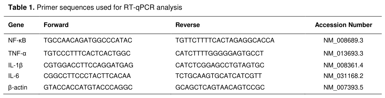
Relative analysis of real-time quantitative PCR gene expressions: RT-qPCR analysis of the target genes NF-κB, TNF-α, IL-1β and IL-6 was performed using the StepOnePlus™ Real-Time PCR System (Applied Biosystems, Foster City, CA, USA) on cDNA synthesized from total RNA isolated from mouse brain tissue. β-actin was employed as an endogenous reference gene in the expression analysis. RT-qPCR was performed using the SYBR Green PCR Master Mix Kit (Ecotech, Erzurum, Türkiye) according to the manufacturer's instructions as previously described. All experiments were performed in triplicate for each sample, and relative gene expression levels were calculated using the 2−ΔΔCT method. The primer sequences are provided in Table 1.
Histopathological examination and semi-quantitative scoring: Brain tissue samples were fixed in 10% neutral-buffered formalin, processed using routine histological procedures, and embedded in paraffin blocks (Paraplast Plus, Sigma-Aldrich, P3683). Serial sections of 5 µm thickness were obtained from the paraffin blocks using a rotary microtome (Leica HistoCore BIOCUT). The sections were stained with hematoxylin and eosin (H&E) to evaluate the general brain tissue morphology and examined under a light microscope (Zeiss Axiolab 5, Jena, Germany) [20].
Histopathological alterations in the cerebral cortex were evaluated using a modified two-component composite semi-quantitative scoring system based on criteria adapted from Kamel et al. [21] and Veldeman et al. [22]. All slides were coded and evaluated by a histologist blinded to the experimental groups. For each animal, five randomly selected, non-overlapping microscopic fields from comparable anatomical regions of the cerebral cortex were examined using a 20× objective. Fields containing tissue folds, edge tears, or prominent tissue-processing artefacts were excluded from the evaluation.
Neuronal degeneration and pericellular vacuolization were evaluated as two separate histopathological components. Degenerated neurons were identified by the combined presence of neuronal cell-body shrinkage or angularity, cytoplasmic hypereosinophilia, and nuclear hyperchromasia or pyknosis. The neuronal damage percentage in each microscopic field was calculated by dividing the number of degenerated neurons by the total number of neurons and multiplying by 100. Pericellular vacuolization was defined as prominent clear spaces immediately surrounding neuronal or neuroglial cell profiles that were not attributable to tissue-processing artefacts. The extent of pericellular vacuolization was evaluated according to the proportion of the examined tissue area affected by this alteration. Perivascular spaces and prominent tissue-processing artefacts were excluded from the assessment.
Both histopathological components were scored separately using the same five-point scale: 0, no lesion; 1, mild involvement affecting >0–25% of the neurons or examined tissue area; 2, moderate involvement affecting >25–50%; 3, severe involvement affecting >50–75%; and 4, extensive involvement affecting >75%. The neuronal degeneration and pericellular vacuolization scores for each microscopic field were summed to obtain a total histopathological injury score ranging from 0 to 8. For each animal, the neuronal degeneration, pericellular vacuolization, and total histopathological injury scores obtained from the five microscopic fields were averaged separately. The mean total score obtained from the five fields was considered the individual total histopathological injury score for that animal. Animals, rather than individual microscopic fields, were considered the experimental units in the statistical analysis. Higher total scores indicated more severe histopathological injury in the cerebral cortex.
Statistical Analysis: Statistical analyses were performed using IBM SPSS Statistics version 30.0 (IBM Corp., Armonk, NY, USA). Graphs were generated using GraphPad Prism version 9.0 (GraphPad Software, San Diego, CA, USA). All data were presented as means ± standard deviation (SD). Prior to analysis, data were tested for normality using the Shapiro–Wilk test and for homogeneity of variance using Levene's test. Comparisons among groups were performed using one-way analysis of variance (ANOVA), followed by Tukey's honestly significant difference (HSD) post hoc test for multiple comparisons. A p value <0.05 was considered statistically significant.
Bulgular
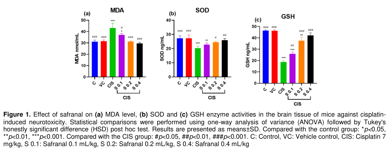
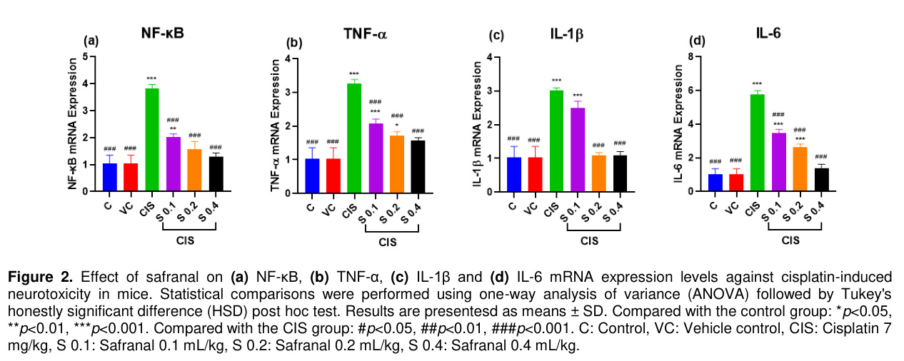
Compared with the control group, the CIS group exhibited significantly higher MDA levels (43.10±4.28 vs. 31.14±1.39, p<0.001) and significantly lower SOD (20.30±1.33 vs. 27.28±1.67, p<0.001) and GSH activities (18.64±1.21 vs. 46.29±0.71, p<0.001). Safranal treatment significantly attenuated CIS-induced oxidative stress. Compared with the CIS group, MDA levels were significantly reduced in the 0.1, 0.2, and 0.4 mL/kg safranal-treated groups (37.10±1.31, p=0.019; 31.22±0.52, p<0.001; and 29.53±1.00, p<0.001, respectively). Likewise, SOD activity was significantly increased in the 0.2 and 0.4 mL/kg groups (24.53±0.73, p=0.021; and 25.95±1.14, p=0.002, respectively), whereas no significant difference was observed in the 0.1 mL/kg group (22.82±1.12, p=0.302). GSH activity was also significantly increased following treatment with 0.1, 0.2, and 0.4 mL/kg safranal (25.86±1.71, p=0.005; 37.43±3.22, p<0.001; and 42.19±2.17, p<0.001, respectively) compared with the CIS group (Figure 1a-c).Compared with the control group, the CIS group exhibited significantly increased mRNA expression levels of NF-κB, TNF-α, IL-1β, and IL-6 (all p<0.001) (Figure 2a-d). No significant differences were observed between the control and vehicle control groups (all p>0.05). Safranal treatment significantly attenuated the CIS-induced upregulation of these inflammatory mediators. Compared with the CIS group, NF-κB, TNF-α, IL-1β, and IL-6 mRNA expression levels were significantly reduced in all safranal-treated groups (all p<0.001). Furthermore, NF-κB mRNA expression in the 0.2 and 0.4 mL/kg safranal groups was comparable to that of the control group (p=0.116 and p=0.741, respectively). Similarly, IL-1β expression in the 0.2 and 0.4 mL/kg groups did not differ significantly from the control group (p=0.999 and p=1.000, respectively). IL-6 expression returned to a level comparable with the control group only in the 0.4 mL/kg safranal group (p=0.620), whereas the 0.2 mL/kg group remained significantly different from the control group (p<0.001). TNF-α expression was reduced in a dose-dependent manner; however, only the 0.4 mL/kg group reached a level comparable to that of the control group (p=0.060), whereas the 0.1 and 0.2 mL/kg groups remained significantly higher than the control group (p<0.001 and p=0.016, respectively).
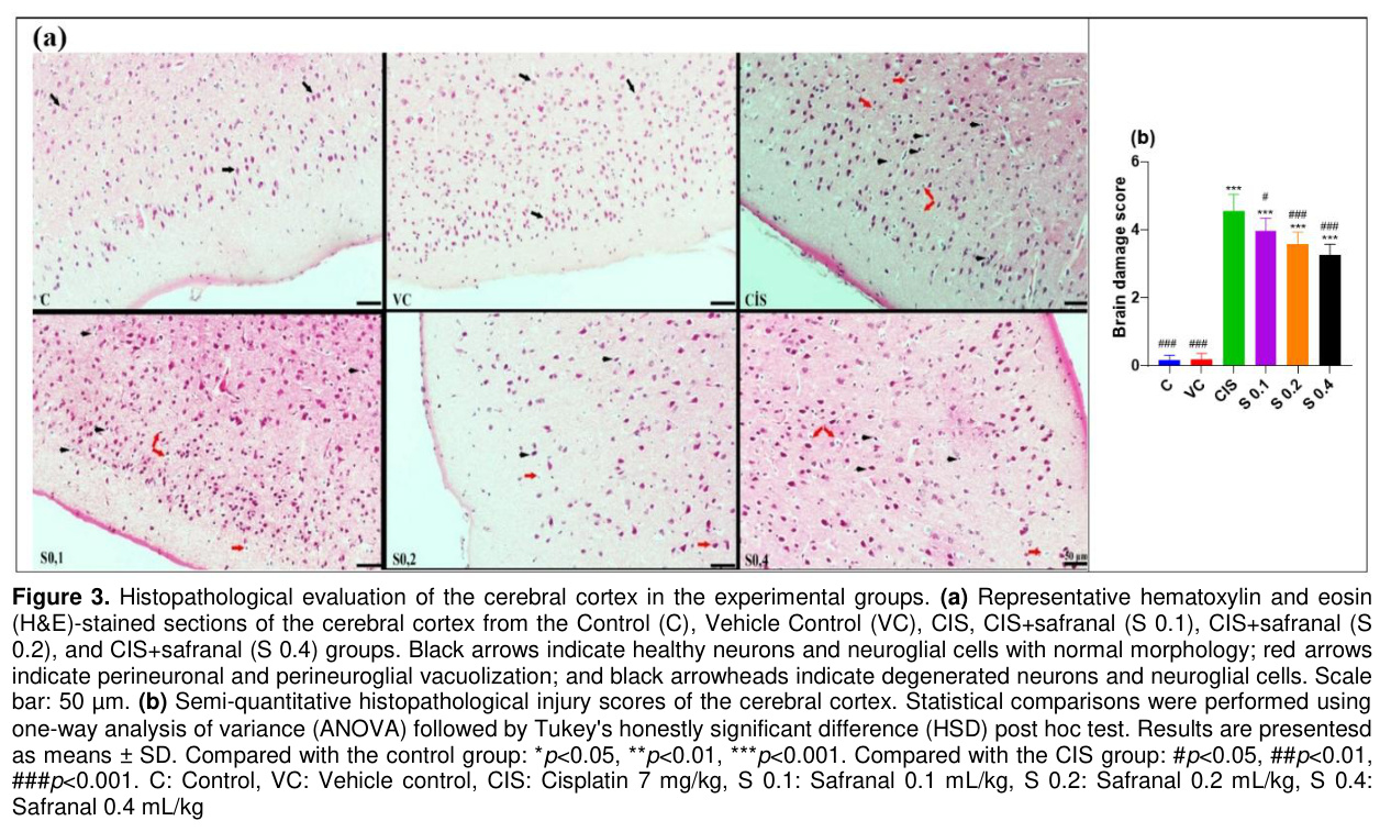
The histomorphological alterations in the cerebral cortex were evaluated using HE staining, as shown in Figure 3a, and the semi-quantitative histopathological injury scores are shown in Figure 3b. The cerebral cortex HE-stained sections were morphologically evaluated at 20× magnification. For each animal, regions selected from the same anatomical area and free of artifacts or edge tears were examined. The evaluation was performed in a blinded manner without knowledge of the experimental groups. During the assessment, the presence and extent of degenerative changes were considered, including hyperchromatic/pyknotic neurons and perineuronal pericellular vacuolization. The findings were descriptively reported based on representative images that best reflected the histopathological of the cerebral cortex in each group. The general histological architecture of the cerebral cortex was preserved in the control group. Evaluation of the control sections revealed that most neurons exhibited normal morphology with no evident degeneration, indicating a normal histological structure representative of the control group. In the vehicle group, the HE appearance of the cerebral cortex was very similar to that of the control group. The neurons predominantly displayed normal morphology with clearly distinguishable nuclear structures, and no marked degenerative changes were observed. Overall, these findings suggest that the administration of the vehicle did not induce significant histopathological alterations. In the CIS group, pronounced degenerative changes were observed in the cortical neurons of the CIS group. An increased number of hyperchromatic/pyknotic neurons was evident in the examined fields. Perineuronal and pericellular vacuolization was more prominent and appeared widespread in certain areas. In the CIS+S 0.1 mL/kg group, partial improvement in tissue integrity was observed compared to that in the CIS-only group; however, degenerative changes were not completely abolished. Hyperchromatic/pyknotic neurons were still present focally, showing similarities to the CIS group. Perineuronal and pericellular vacuolization was mild to moderate and became more evident in some regions. In the CIS+S 0.2 mL/kg group, the cortical structure appeared better preserved than that in the 0.1 mL/kg safranal group. Hyperchromatic/pyknotic neurons were observed less frequently, and perineuronal and pericellular vacuolization were minimal. In the CIS+S 0.4 mL/kg group, the histomorphology of the cerebral cortex was largely preserved and exhibited an appearance similar to that of the control/sham groups. Hyperchromatic/pyknotic neurons were minimal and not widespread. Although mild perineuronal/pericellular vacuolization was observed, it was markedly reduced compared to that in the CIS group. The semi-quantitative histopathological injury scores (Figure 3b) supported the histopathological findings. The CIS group exhibited a significantly higher histopathological injury score than the control group (p<0.001). No significant difference was observed between the control and vehicle groups (p>0.05). Compared with the CIS group, safranal treatment significantly reduced the histopathological injury score at doses of 0.1 mL/kg (p<0.05), 0.2 mL/kg (p<0.001), and 0.4 mL/kg (p<0.001).
Tartışma
Cisplatin is a chemotherapeutic agent used to treat brain, mammary and lung cancer. Its effectiveness is dose dependent, showing better results at higher doses. However, it causes adverse effects including neurotoxicity, cardiotoxicity, nephrotoxicity and gastrointestinal toxicity [23,24]. These effects limit cisplatin's administration at effective doses and impact patients' quality of life. No official prophylactic cautions have been determined to neutralize cisplatin's neurotoxic effects [5]. Neurotoxicity is one of the most important side effects in cisplatin's clinical use, commonly addressed through dose modifications and treatment discontinuation [25-27]. Safranal is an aromatic component extracted from the saffron plant (Crocus sativus) that has pharmacologic activities such as antioxidant and anti-inflammatory effects [11,25]. In the present study, we found that safranal treatment attenuated CIS-induced neurotoxicity in mice, as evidenced by improvements in oxidative stress, inflammatory markers, and histopathological alterations.Oxidative stress is characterized by the disruption of the physiological redox balance, resulting in decreased endogenous antioxidants such as SOD, CAT, GSH, and GSH-Px, together with increased oxidative biomarkers, including MDA [28-31]. Neuronal cells are particularly susceptible to oxidative stress because of their high lipid content and limited antioxidant capacity, making them more vulnerable to oxidative damage. Consequently, oxidative stress may induce structural alterations in neural tissue through demyelination and myelin degeneration [32]. Oxidative stress is considered one of the principal mechanisms underlying CIS-induced neurotoxicity [33]. In the present study, CIS exposure significantly increased MDA levels while decreasing SOD and GSH activities in mouse brain tissue, confirming the induction of oxidative stress. Consistent with previous studies [34], safranal treatment dose-dependently attenuated these alterations, as evidenced by reduced MDA levels and restoration of SOD and GSH activities, suggesting that it may preserve the endogenous antioxidant defense system. Similar protective effects have also been reported following gossypin treatment in experimental models of CIS-induced neurotoxicity [5], supporting the importance of antioxidant strategies in limiting neuronal damage.
In addition to cellular oxidative stress, neuro-inflammation is another possible mechanism involved in CIS-induced toxicity. Increased levels of reactive oxygen species, pro-inflammatory cytokines, and inflammatory mediators indicate neuronal toxicity that causes cognitive deficits through cell death or damage [34]. CIS triggers the release of TNF-α, which crosses the blood-brain barrier via receptor-mediated endocytosis, causing neuro-oxidative damage. CIS also accumulates in mitochondria, leading to an increase in ROS and pro-inflammatory molecules. Therefore, compounds that reduce oxidative stress and inflammation may offer potential therapeutic strategies against chemotherapy-induced cerebellar damage [21,35,36]. Safranal has been reported to have anti-inflammatory activities against various toxic substances [37]. Many studies have established that CIS increases the transcription factor NF-κB in brain tissue, triggering the transcription of pro-inflammatory cytokines such as TNF-α, IL-1β, and IL-6, thereby initiating and sustaining an inflammatory response. Several studies have suggested that TNFα is a key regulator of inflammatory activities after CIS exposure and is responsible for the elevated levels of inflammatory cytokines and chemokines [38]. A previous study also reported that gossypin treatment downregulated the expression of NF-κB and pro-inflammatory cytokines (TNFα, IL1-β and IL-6) in a CIS-induced neurotoxicity model [5]. In this study, consistent with previous studies, we observed significantly higher levels of TNFα, IL-1β and IL-6 in brain tissue of mice exposed to CIS, suggesting a role for inflammatory cytokines in neurotoxicity. Interestingly, the levels of these inflammatory cytokines significantly decreased after safranal pre-treatment, consistent with a potential neuroprotective effect of safranal against cisplatin CIS-induced neurotoxicity.
Several plant-derived extracts and phytochemicals have been investigated as potential neuroprotective agents against cisplatin-induced neurotoxicity because of their antioxidant and anti-inflammatory properties [8]. For example, Ginkgo biloba extract preserved neuronal integrity by scavenging reactive oxygen and nitrogen species [39], whereas curcumin alleviated oxidative stress, improved mitochondrial function, and reduced histopathological damage [40]. Similarly, thymoquinone enhanced endogenous antioxidant defenses and suppressed oxidative injury [41], while extracts of Agrimonia eupatoria [42], Salvia officinalis [43], Matricaria chamomilla [44], and alkaloids isolated from Corydalis saxicola [45] reduced the production of pro-inflammatory mediators, including TNF-α, IL-1β, IL-6, and PGE₂. Collectively, these findings indicate that attenuation of oxidative stress and neuroinflammation represents a common mechanism underlying the neuroprotective effects of plant-derived compounds against cisplatin-induced neurotoxicity. Consistent with these observations, safranal treatment in the present study significantly reduced oxidative stress, suppressed NF-κB, TNF-α, IL-1β and IL-6 expression, and improved histopathological alterations in brain tissue. These findings suggest that the neuroprotective effects of safranal may involve mechanisms similar to those reported for other plant-derived compounds. Gossypin normalized SOD and GSH levels while suppressing NF-κB and TNF-α expression [5]. Likewise, mesna [6], N-acetylcysteine [46], agmatine [47], and calcitriol [48] have all been reported to reduce oxidative stress and inflammatory responses in experimental models of CIS-induced neurotoxicity. Similar biochemical and molecular changes observed following safranal treatment further support its potential neuroprotective profile.
Collectively, these previous studies together with our findings suggest that modulation of oxidative stress and inflammatory pathways may represent a common mechanism underlying protection against CIS-induced neurotoxicity. In the present study, safranal improved biochemical, molecular, and histopathological outcomes, supporting its potential as a promising experimental candidate for reducing cisplatin-induced neurotoxicity. Nevertheless, further mechanistic investigations and well-designed clinical studies are required before its clinical application can be considered.
References
- Bilgiç S, Aktaş İ, Yahyazadeh A. Protection of lutein against the neurotoxicity of cisplatin in the rat brain. Tissue Cell 2024; 91:102609.
- Huang Y, Liu C, Song X, et al. Antioxidant and anti-inflammatory properties mediate the neuroprotective effects of hydro-ethanolic extract of Tiliacora triandra against cisplatin-induced neurotoxicity. J Inflamm Res 2021; 14: 6735-6748.
- Li Y, Squillace S, Schafer R, et al. Contribution of S1pr1-featured astrocyte subpopulation to cisplatin-induced neuropathic pain in male mice. Pain 2025; 167(1): 204-217.
- Gupta P, Makkar TK, Goel L, Pahuja M. Role of inflammation and oxidative stress in chemotherapy-induced neurotoxicity. Immunol Res 2022; 70(6): 725-741.
- Cinar I, Yayla M, Bayram P. Comparison of the neuroprotective effects of gossypin on cisplatin-induced neurotoxicity in vitro and in vivo. Mol Biol Rep 2025; 52(1): 412.
- Saadati H, Noroozzadeh S, Esmaeili H, et al. The neuroprotective effect of mesna on cisplatin-induced neurotoxicity: Behavioral, electrophysiological, and molecular studies. Neurotox Res 2021; 39 (3): 826-840.
- Fazeli E, Ghalibaf MHE, Forouzanfar F. Neuroprotective potency of safranal against neurological disorders. Curr Mol Med 2023; 23(9): 952-959.
- Xu X, Jia L, Ma X, Li H, Sun C. Application potential of plant-derived medicines in prevention and treatment of platinum-induced peripheral neurotoxicity. Front Pharmacol 2022; 12: 792331.
- Karafakıoğlu YS, Bozkurt MF, Hazman Ö, Fidan AF. Efficacy of safranal to cisplatin-induced nephrotoxicity. Biochem J 2017; 474(7): 1195-1203.
- Yan J, Li T, Ji K, et al. Safranal alleviates pentetrazole-induced epileptic seizures in mice by inhibiting the NF-κB signaling pathway and mitochondrial-dependent apoptosis through GSK-3β inactivation. J Ethnopharmacol 2024; 333: 118408.
- Cellat M, Yavaş İ, Uyar A, et al. Safranal alleviates cyclophosphamide-induced testicular toxicity in rats. Food Sci Nutr 2025; 13 (6): e70452.
- Etyemez M, Gülay MŞ. The effects of safranal against bisphenol AF on some reproductive parameters in male New zealand rabbits. Vet J Mehmet Akif Ersoy Univ 2022; 7 (3): 207-217.
- Cerda-Bernad D, Valero-Cases E, Pastor JJ, Frutos MC. Saffron bioactives crocin, crocetin and safranal: Effect on oxidative stress and mechanisms of action. Crit Rev Food Sci Nutr 2022; 62 (12): 3232-3249.
- Baluchnejadmojarad T, Mohamadi-Zarch S-M, Roghani M. Safranal, an active ingredient of saffron, attenuates cognitive deficits in amyloid β-induced rat model of Alzheimer's disease: Underlying mechanisms. Metab Brain Dis 2019; 34 (6): 1747-1759.
- Gupta M, Wani A, Ahsan AU, et al. Safranal inhibits NLRP3 inflammasome activation by preventing ASC oligomerization. Toxicol Appl Pharmacol 2021; 423: 115582.
- Hosseinzadeh H, Sadeghnia H. Protective effect of safranal on pentylenetetrazol-induced seizures in the rat: Involvement of GABAergic and opioids systems. Phytomedicine 2007; 14 (4): 256-262.
- Amin B, Malekzadeh M, Heidari MR, et al. Effect of Crocus sativus extracts and its active constituent safranal on the harmaline-induced tremor in mice. Iran J Basic Med Sci 2015; 18(5): 449-458.
- Ibrahim MA, Bakhaat GA, Tammam HG, Mohamed RM, El-Naggar SA. Cardioprotective effect of green tea extract and vitamin E on Cisplatin-induced cardiotoxicity in mice: Toxicological, histological and immunohistochemical studies. Miomed Pharmacother 2019; 113: 108731.
- Yang W, Wei Y, Sun J, et al. Safranal exerts a neuroprotective effect on Parkinson’s disease with suppression of NLRP3 inflammation activation. Mol Biol Rep 2024; 51 (1): 593.
- Bancroft JD, Gamble M. Theory and Practice of Histological Techniques. Elsevier Health Sciences; 2008.
- Kamel NA, Bashir DW, El-Leithy EMM, et al. Polyethylene terephthalate nanoplastics-induced neurotoxicity in adult male Swiss albino mice with amelioration of betaine: A histopathological, neurochemical, and molecular investigation. Naunyn Schmiedeberg's Arch Pharmacol 2025; 398(7): 9323-9339.
- Veldeman M, Coburn M, Rossaint R, et al. Xenon reduces neuronal hippocampal damage and alters the pattern of microglial activation after experimental subarachnoid hemorrhage: A randomized controlled animal trial. Front Neurol 2017; 8: 511.
- Tchounwou PB, Dasari S, Noubissi FK, et al. Advances in our understanding of the molecular mechanisms of action of cisplatin in cancer therapy. J Exp Pharmacol 2021; 13: 303-328.
- Altunkaya M, Ateş MB, Bulut A, et al. Cisplatin-induced toxicity in the hippocampus: A dose-dependent mechanism of damage. BMC Pharmacol Toxicol 2025; 26(1): 215.
- Was H, Borkowska A, Bagues A, et al. Mechanisms of chemotherapy-induced neurotoxicity. Front Pharmacol 2022; 13: 750507.
- Tao Z, Chen Z, Zeng X, et al. An emerging aspect of cancer neuroscience: A literature review on chemotherapy-induced peripheral neuropathy. Cancer Lett 2025; 611: 217433.
- Ishak L, Jasaputra DK, Gunadi JW, Samuel LM, Rumanti RT. The potential role of saffron extract, crocin, and crocetin in modulating TNF-α and IL-6 levels in various diseases: A review. Trop J Nat Prod Res 2025; 9(2): 424-431.
- Lin SY, Chang CL, Liou KT, et al. The protective role of Achyranthes aspera extract against cisplatin-induced nephrotoxicity by alleviating oxidative stress, inflammation, and PANoptosis. J Ethnopharmacol 2024; 319: 117097.
- Bademci R, Erdoğan MA, Eroğlu E, et al. Demonstration of the protective effect of ghrelin in the livers of rats with cisplatin toxicity. Hum Exp Toxicol 2021; 40(12): 2178-2187.
- Issı M, Gul Y, Yılmaz S. Clinical, haematological and antioxidant status in naturally poxvirus infected sheep. Revue Méd Vét 2008; 159(1): 54-58.
- Yılmaz S, Issı M, Kandemır FM, Gul Y. Malondialdehyde and total antioxidant levels and hematological parameters of beef cattle with coccidiosis. Van Veterinary Journal 2014; 25(2): 41-45.
- Dash UC, Bhol NK, Swain SK, et al. Oxidative stress and inflammation in the pathogenesis of neurological disorders: Mechanisms and implications. Acta Pharm Sin B 2025; 15(1): 15-34.
- Attia A, Matta C, ElMazoudy R, et al. Cisplatin-induced neurotoxicity in cerebellar cortex of male mice involves oxidative stress and histopathology. J Basic Appl Zool 2021; 82(1): 23.
- Checa J, Aran JM. Reactive oxygen species: Drivers of physiological and pathological processes. J Inflamm Res 2020; 13: 1057-1073.
- Cocetta V, Ragazzi E, Montopoli M. Mitochondrial involvement in cisplatin resistance. Int J Mol Sci 2019; 20(14): 3384.
- Seo DY, Bae JH, Zhang D, et al. Effects of cisplatin on mitochondrial function and autophagy-related proteins in skeletal muscle of rats. BMB Rep 2021; 54(11): 575-580.
- Nanda S, Madan K. The role of Safranal and saffron stigma extracts in oxidative stress, diseases and photoaging: A systematic review. Heliyon 2021; 7(2): e06117.
- Anwer T, Alshahrani S, Somaili AM, et al. Nephroprotective effect of diosmin against cisplatin-induced kidney damage by modulating IL-1β, IL-6, TNFα and renal oxidative damage. Molecules 2023; 28(3): 1302.
- Diamond BJ, Shiflett SC, Feiwel N, et al. Ginkgo biloba extract: mechanisms and clinical indications. Arch Phys Med Rehabil 2000; 81(5): 668-678.
- Agthong S, Kaewsema A, Charoensub T. Curcumin ameliorates functional and structural abnormalities in cisplatin-induced neuropathy. Exp Neurobiol 2015; 24(2): 139-145.
- Üstün R, Oğuz EK, Şeker A, Korkaya H. Thymoquinone prevents cisplatin neurotoxicity in primary DRG neurons. Neurotoxicology 2018; 69: 68-76.
- Lee KH, Rhee KH. Anti-nociceptive effect of Agrimonia eupatoria extract on a cisplatin-induced neuropathic model. Afr J Tradit Complement Altern Med 2016; 13(5): 139-144.
- Namvaran-Abbas-Abad A, Tavakkoli F. Antinociceptive effect of Salvia extract on cisplatin-induced hyperalgesia in mice. Neurophysiology 2012; 43(6): 452-458.
- Abad ANA, Nouri MHK, Gharjanie A, Tavakoli F. Effect of Matricaria chamomilla hydroalcoholic extract on cisplatin-induced neuropathy in mice. Chin J Nat Med 2011; 9(2): 126-131.
- Kuai CP, Ju LJ, Hu PP, Huang F. Corydalis saxicola alkaloids attenuate cisplatin-induced neuropathic pain by reducing loss of IENF and blocking TRPV1 activation. Am J Chin Med 2020; 48(2): 407-428.
- Abdel-Wahab WM, Moussa FI. Neuroprotective effect of N-acetylcysteine against cisplatin-induced toxicity in rat brain by modulation of oxidative stress and inflammation. Drug Des Devel Ther 2019; 13: 1155-1162.
- Binnetoglu D, Hacimuftuoglu A, Aricioglu F. Neuroprotective effects of agmatine in antineoplastic drugs induced neurotoxicity: In vitro study. Life Sci 2019; 221: 311-318.
- Niapour A, Abdollahzadeh M, Ghaheri Fard S, et al. The therapeutic potential of 1, 25-dihydroxy vitamin D3 on cisplatin-affected neurological functions is associated with the regulation of oxidative stress and inflammatory markers as well as levels of MMP2/9. Metab Brain Dis 2024; 39(6): 1189-1200.
- ŞAP AŞISI UYGULANAN BESİ SIĞIRLARINDA ANTİOKSİDAN VİTAMİNLERİN KLİNİK VE BAZI HEMATOLOJİK PARAMETRELER İLE ANTİOKSİDAN ENZİM VE LİPİT PEROKSİDASYON DÜZEYLERİNE ETKİLERİ
- Lenfatik Uterus Kistli Arap Kısraklarında Plazma Oxidan-Antioxidant Değerleri
- Ratların Karaciğer ve Testis Dokusundaki Antioksidan Aktivite Üzerine Nar Suyunun Etkisi
- Yüksek Enerjili Diyetler ve Soğuk Çevre Şartlarında (15 ºC) Yetiştirilen Broylerlerde Selenyum ve Vitamin C Katkısının Lipit Peroksidasyon Üzerine Etkileri
- Elektromanyetik Radyasyona Maruz Bırakılan Kobayların Böbrek Dokusunda Prooksidan-Antioksidan Düzeylerine Yeşil Çayın Etkisi