
Koyun Somatik Hücre Nükleer Transferinde Konvansiyonel ve Piezo Destekli Oosit Rekonstrüksiyon Yöntemlerinin Karşılaştırılması
Cilt 40, Sayı 3
Ekim 2026 · Sayfalar 266-272
Koyun Somatik Hücre Nükleer Transferinde Konvansiyonel ve Piezo Destekli Oosit Rekonstrüksiyon Yöntemlerinin Karşılaştırılması
Comparison of Conventional and Piezo-Assisted Oocyte Reconstruction Methods for Ovine Somatic Cell Nuclear Transfer
Selin YAĞCIOĞLU1, Ramazan ARICI1, Gül BAKIRER ÖZTÜRK2, Ahmet ESER3, Kamber DEMİR1, Mithat EVECEN1, Sema BİRLER1, Serhat PABUCCUOĞLU1
2İstanbul University, Aziz Sancar Institute of Experimental Medicine, Department of Laboratory Animal Sciences, İstanbul, TÜRKİYE
3Siirt University, Faculty of Veterinary Medicine, Department of Reproduction and Artificial Insemination, Siirt, TÜRKİYE
Somatik hücre nükleer transferi (SCNT), çiftlik hayvanlarında kullanılan yardımcı üreme teknolojileri arasında yer almakla birlikte, embriyo rekonstrüksiyonu sırasında meydana gelen teknik kayıplar klonlama verimliliğini sınırlayan önemli bir faktör olmaya devam etmektedir. Bu çalışmada, koyunlarda konvansiyonel SCNT yöntemi ile karşılaştırıldığında piezo destekli oosit rekonstrüksiyonunun rekonstrüksiyon aşamasındaki teknik performansı ve embriyo gelişimini iyileştirip iyileştirmediği değerlendirilmiştir. İn vitro olgunlaştırılan oositler enüklee edildikten sonra, rekonstrükte oositler ya donör hücrenin perivitellin boşluğa yerleştirilmesini takiben elektrofüzyon uygulanarak ya da piezo destekli doğrudan intrasitoplazmik donör hücre transferi ile oluşturulmuştur. Enükleasyon başarısı yöntemler arasında farklılık göstermezken (p=0.112), rekonstrüksiyon başarısı piezo destekli grupta konvansiyonel gruba göre sınırlı ancak anlamlı düzeyde daha yüksek bulunmuştur (%98.5'e karşı %96.8; p=0.044). Piezo destekli rekonstrüksiyon sağlam rekonstrükte oosit oranını anlamlı düzeyde artırmış (%73.3'e karşı %51.8) ve oosit lizisini azaltmıştır (%26.7'ye karşı %48.2; p<0.001). Ayrıca bu yöntem, bölünme (%30.5'e karşı %22.4; p=0.015), toplam embriyo gelişimi (%46.7'ye karşı %35.5; p=0.002) ve pre-morula gelişimi (%16.6'ya karşı %9.0; p=0.003) oranlarını artırırken, dejenere embriyo oranını anlamlı düzeyde azaltmıştır (%13.2'ye karşı %27.8; p<0.001). Morula–erken blastosist gelişimi bakımından gruplar arasında anlamlı bir farklılık saptanmamıştır (p=0.841). Bu bulgular, piezo destekli rekonstrüksiyonun oosit bütünlüğünü daha iyi koruduğunu, manipülasyona bağlı hasarı azalttığını ve erken embriyonik gelişimi iyileştirdiğini göstermekte olup, koyun SCNT uygulamalarında verimliliğin artırılmasına yönelik uygulanabilir bir strateji sunduğunu ortaya koymaktadır.
Although somatic cell nuclear transfer (SCNT) is among the assisted reproductive technologies utilized in livestock, technical losses occurring during embryo reconstruction remain one of the significant factors limiting cloning efficiency. This study evaluated whether piezo-assisted oocyte reconstruction could improve reconstruction-stage technical performance and embryonic development compared with the conventional SCNT method in sheep. In vitro-matured oocytes were enucleated and reconstructed by either conventional perivitelline donor cell placement followed by electrofusion or direct piezo-assisted intracytoplasmic donor cell transfer. Enucleation success did not differ between methods (p=0.112), whereas reconstruction success was modestly but significantly higher in the piezo-assisted group (98.5% vs. 96.8%; p=0.044). Piezo-assisted reconstruction also significantly increased the proportion of intact reconstructed oocytes (73.3% vs. 51.8%) and reduced oocyte lysis (26.7% vs. 48.2%; p<0.001). It also improved cleavage (30.5% vs. 22.4%; p=0.015), overall embryo development (46.7% vs. 35.5%; p=0.002), and pre-morula development (16.6% vs. 9.0%; p=0.003), while significantly reducing embryo degeneration (13.2% vs. 27.8%; p<0.001). Morula–early blastocyst development was similar between groups (p=0.841). These findings demonstrate that piezo-assisted reconstruction better preserves oocyte integrity, reduces manipulation-induced damage, and enhances early embryonic development, representing a practical strategy for improving ovine SCNT efficiency.
Giriş
Somatic cell nuclear transfer (SCNT) is an assisted reproductive technology in which the nucleus of a differentiated somatic cell is transferred into an enucleated metaphase II oocyte to generate a reconstructed embryo. The successful birth of Dolly, the first mammal cloned from an adult somatic cell in 1997, demonstrated that differentiated somatic nuclei can be reprogrammed by the oocyte cytoplasm to support full-term development. This landmark achievement established sheep as a model for mammalian cloning and represented a major milestone in developmental biology and reproductive biotechnology [1]. Since the birth of Dolly, SCNT has become an important tool for livestock breeding, production of transgenic animals, conservation of endangered species, and biomedical research. More recently, its integration with genome-editing technologies has further expanded its applications [2,3].The developmental competence of cloned embryos is influenced by multiple biological and technical factors throughout the SCNT procedure. Successful cloning depends on high-quality oocytes, accurate enucleation, efficient embryo reconstruction, appropriate activation, and optimal embryo culture. Although incomplete epigenetic reprogramming remains a major limitation, refinement of technical procedures, particularly embryo reconstruction and micromanipulation techniques, is also essential for improving developmental competence [2,4]. In sheep, despite continuous methodological improvements in donor cell preparation, embryo reconstruction, and activation protocols, cloning efficiency remains relatively low [5,6]. Among the reconstruction procedures, donor cell transfer represents one of the most delicate micromanipulation steps. Conventionally, donor cells are introduced into the perivitelline space and fused with the recipient cytoplast using electrical pulses. Although routinely employed in livestock cloning, electrofusion may cause incomplete membrane fusion, membrane disruption, oocyte lysis, and degeneration of reconstructed oocytes [7-9]. To overcome these limitations, piezo-assisted reconstruction has been introduced as an alternative approach in which the donor cell is delivered directly into the ooplasm, thereby eliminating the need for electrofusion. Previous studies have suggested that piezo-assisted reconstruction may improve reconstruction-stage technical performance by reducing mechanical manipulation and minimizing oocyte damage, although its effects on subsequent embryo development appear to depend on manipulation parameters and experimental conditions [7,10]. Nevertheless, comparative evaluations of conventional and piezo-assisted reconstruction during ovine SCNT remain limited [11].
Although previous ovine SCNT studies have mainly focused on embryo development and cloning efficiency, reconstruction-stage technical performance has received comparatively little attention [11,12]. The present study aimed to compare conventional and piezo-assisted reconstruction methods during ovine SCNT, with particular emphasis on reconstruction-stage technical performance, including reconstruction success, post-manipulation oocyte integrity, oocyte lysis, and subsequent embryonic development.
Gereç ve Yöntem
Research and Publication Ethics: The experimental procedures were approved by the Istanbul University Animal Experiments Local Ethics Committee (Approval No. 2016/33; 30 June 2016). Ovaries and donor tissues were collected from sheep slaughtered for commercial purposes at a local abattoir.Experimental Design: The study compared two embryo reconstruction methods used during ovine somatic cell nuclear transfer (SCNT): (i) conventional perivitelline donor cell placement followed by electrofusion and (ii) piezo-assisted intracytoplasmic donor cell transfer. Recipient oocytes in both groups underwent identical procedures for in vitro maturation, enucleation, chemical activation, and embryo culture; the only experimental variable was the reconstruction method. The study was conducted over 12 independent experimental sessions. Within each session, oocytes processed under the same laboratory conditions were allocated between the conventional and piezo-assisted reconstruction groups, and both reconstruction procedures were performed in parallel. Reconstructed oocytes from both groups were subsequently cultured under identical conditions within the corresponding experimental session. Oocytes recovered from slaughterhouse-derived ovaries were pooled during collection and processing; therefore, individual oocytes could not be traced to a specific donor animal or ovary.
Reconstruction-stage technical performance was assessed based on reconstruction success, post-manipulation oocyte integrity, and oocyte lysis. Embryonic development was evaluated by cleavage, overall embryo development, non-cleaved embryos, pre-morula formation, morula–early blastocyst development, embryo fragmentation, and embryo degeneration following in vitro culture.
Preparation and Synchronization of Donor Fibroblasts: Primary adult ovine fibroblasts were established from ear skin collected immediately after slaughter at a local abattoir. Tissue samples were transported to the laboratory in phosphate-buffered saline (PBS) supplemented with antibiotics. After removal of cartilage and subcutaneous connective tissue, skin explants were cultured in 60-mm dishes containing Dulbecco's Modified Eagle Medium (DMEM) supplemented with 20% fetal bovine serum (FBS) and 1% antibiotic-antimycotic solution. Cultures were maintained at 38.5°C in a humidified atmosphere of 5% CO₂ until fibroblasts reached approximately 70% confluence. Cells were detached with 0.25% trypsin-EDTA, subcultured, and cryopreserved at passages 2–3 in freezing medium containing 20% FBS and 10% dimethyl sulfoxide (DMSO). Prior to SCNT, donor fibroblasts were synchronized by contact inhibition. Cells were maintained at complete confluence for an additional 5 days in DMEM containing 10% FBS to induce G0/G1 synchronization, as previously described by Yagcioglu et al. [13].
Oocyte Collection, in vitro Maturation and Selection of Oocytes: Sheep ovaries were collected from a local abattoir and transported to the laboratory in PBS maintained at 30–35°C. Cumulus–oocyte complexes (COCs) were recovered by the ovarian slicing method in oocyte washing medium (OWM). Only Grade I and II COCs with at least three to four compact cumulus cell layers and homogeneous ooplasm were selected for in vitro maturation. Selected COCs were matured in commercial BO-IVM medium (IVF Bioscience, Falmouth, UK) at 38.5°C in a humidified atmosphere of 5% CO₂ for 20 h. Cumulus cells were then removed by vortexing in HEPES-buffered synthetic oviductal fluid (SOF) medium containing 300 IU/mL hyaluronidase, and only metaphase II oocytes exhibiting the first polar body were selected for SCNT. Oocyte collection, in vitro maturation, and oocyte selection were performed as previously described, with minor modifications [5,6].
Enucleation and Embryo Reconstruction: Metaphase II (MII) oocytes were randomly allocated to either the conventional or piezo-assisted reconstruction group prior to enucleation. Oocytes in both groups underwent the same enucleation procedure and were incubated in manipulation medium containing 5 µg/mL Hoechst 33342 for 15 min and then transferred to enucleation medium supplemented with 7.5 µg/mL cytochalasin B. Enucleation was performed under an inverted microscope (Olympus IX71, Olympus Optical Co., Tokyo, Japan) equipped with Eppendorf micromanipulators (TransferMan NK2; Eppendorf AG, Hamburg, Germany) by aspirating the first polar body together with the adjacent metaphase II spindle. Custom-made holding (120–130 µm outer diameter) and donor cell transfer pipettes (20 µm inner diameter; spiked- or straight-tip), fabricated from borosilicate glass capillaries, were used for all micromanipulation procedures. Enucleated oocytes (cytoplasts) were subsequently incubated in SOF medium for 30 min before donor cell transfer. Following enucleation, embryo reconstruction was performed according to the previously assigned group, either by conventional perivitelline donor cell placement followed by electrofusion or by piezo-assisted direct intracytoplasmic donor cell transfer. In both groups, a single contact inhibition-synchronized adult fibroblast served as the donor cell, and reconstructed oocytes subsequently underwent identical chemical activation [5,6].
Conventional SCNT Reconstruction: In the conventional SCNT group, a single donor fibroblast was placed into the perivitelline space using a spiked donor cell transfer pipette. Donor cell–cytoplast complexes were transferred into fusion medium consisting of 0.3 M mannitol, 0.1 mM MgCl₂ and 0.05 mM CaCl₂. Cell fusion was induced using a single direct-current pulse of 1.25 kV/cm for 60–80 µs. Reconstructed oocytes were incubated for 30–45 min under standard culture conditions, after which successful fusion was assessed microscopically [5,6].
Piezo-assisted SCNT Reconstruction: In the piezo-assisted group, a single donor fibroblast was aspirated into a straight-tip borosilicate injection pipette with an inner diameter of 15 µm and an outer diameter of 20 µm. Mercury was used as the heavy liquid in the injection pipette. Piezo-assisted manipulation was performed using a PMM-4G piezo-assisted micromanipulation system (Prime Tech Ltd., Ibaraki, Japan) operated in Expert mode. The donor cell was repeatedly aspirated and expelled within the injection pipette while one or two piezo pulses were applied using Condition B (intensity 8, speed 7) to induce plasma membrane permeabilization. The injection pipette was then advanced through the zona pellucida and oolemma. A single piezo pulse was applied for zona pellucida penetration using Condition B (intensity 8, speed 7), whereas a single piezo pulse was applied for oolemma penetration using Condition A (intensity 4, speed 2). The injection site was selected in a region with a clearly visible perivitelline space between the zona pellucida and the oolemma to minimize mechanical damage to the oocyte membrane. Following oolemma penetration, the donor cell was deposited directly into the ooplasm.
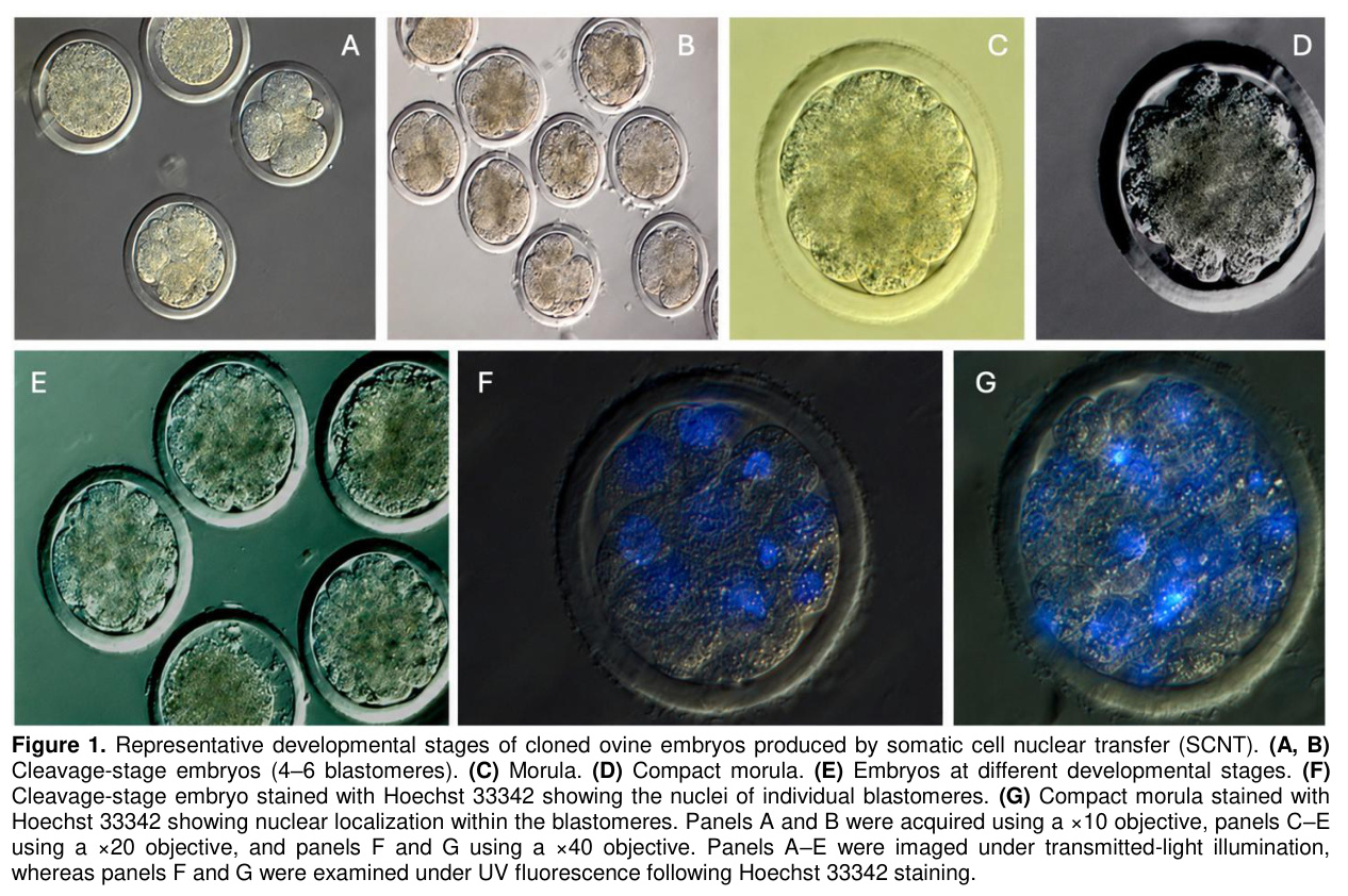
Oocyte Activation and Embryo Culture: Following donor cell transfer, reconstructed oocytes in both groups were chemically activated by incubation in 5 µg/mL calcium ionophore for 5 min, followed by culture in 6-dimethylaminopurine (6-DMAP) for 3 h. Activated embryos were washed and cultured in BO-IVC medium (IVF Bioscience, Falmouth, UK) at 38.5°C in a humidified atmosphere of 5% CO₂, 5% O₂, and 90% N₂. Cleavage was evaluated at 48 h post-activation, whereas further embryonic development was evaluated on Day 8 of culture [6]. For fluorescence evaluation, representative embryos were fixed in ice-cold ethanol containing 10 µg/mL Hoechst 33342 for 15 min and subsequently examined under UV illumination using an Olympus IX71 fluorescence microscope (Olympus, Tokyo, Japan).
Evaluation of Reconstruction-stage Technical Performance: The technical performance of the two reconstruction methods was evaluated by assessing reconstruction success, post-manipulation oocyte integrity, and oocyte lysis. Reconstruction success was defined as complete donor cell–cytoplast fusion following electrofusion in the conventional group and successful intracytoplasmic deposition of the donor fibroblast immediately after manipulation in the piezo-assisted group. Following reconstruction, oocytes with an intact oolemma and normal cytoplasmic morphology were classified as intact reconstructed oocytes, whereas those showing membrane rupture, cytoplasmic leakage, or degeneration were classified as lysed oocytes. Following in vitro culture, embryonic development was assessed at defined time points. Cleavage was evaluated at 48 h post-activation. Embryos showing blastomere division were classified as cleaved embryos, whereas fragmented embryos were recorded as a separate developmental outcome and were not included in the cleavage category. Overall embryo development included both cleaved and fragmented embryos. Further embryonic development was evaluated on Day 8 of culture. Embryos that showed no cleavage but did not meet the criteria for degeneration were classified as non-cleaved embryos, whereas embryos showing morphological degeneration were classified as degenerated embryos. On Day 8, cleaved embryos were further classified as pre-morula or morula–early blastocyst according to their morphological stage. The morula–early blastocyst category included embryos ranging from the morula and compacted morula stages to morphologically identified early blastocysts. Percentages for developmental outcomes were calculated relative to the total number of reconstructed embryos entering in vitro culture within each reconstruction group.
Statistical Analysis: Statistical analyses were performed using IBM SPSS Statistics version 21.0 (IBM Corp., Armonk, NY, USA). Categorical data, including enucleation success, reconstruction success, oocyte integrity, oocyte lysis, cleavage, overall embryo development, non-cleaved embryos, pre-morula formation, morula–early blastocyst development, embryo fragmentation, and embryo degeneration, were expressed as frequencies and percentages. Differences between the conventional and piezo-assisted reconstruction groups were analyzed using Pearson's Chi-square test. Individual oocytes and embryos were used as observational units for these frequency-based comparisons, whereas the 12 experimental sessions represented the independent experimental replicates. Because oocytes recovered from slaughterhouse-derived ovaries were pooled during collection and processing, individual oocytes could not be traced to a specific donor animal or ovary; therefore, clustering at the donor-animal or ovary level could not be incorporated into the statistical analysis. Expected cell frequencies were examined and were adequate for the application of Pearson's Chi-square test in all comparisons. The final sample size reflected all eligible oocytes available and processed across the 12 experimental sessions, and no prospective sample-size calculation was performed. A two-tailed p<0.05 was considered statistically significant. Exact p values are reported where appropriate, whereas values below 0.001 are reported as p<0.001.
Bulgular
Technical Performance of Reconstruction Methods: A total of 1,727 metaphase II oocytes were used for SCNT, of which 915 were allocated to the conventional reconstruction group and 812 to the piezo-assisted reconstruction group (Table 1). Successful enucleation was achieved in 654/915 (71.5%) and 608/812 (74.9%) oocytes in the conventional and piezo-assisted groups, respectively. Of the successfully enucleated oocytes, 633/654 (96.8%) and 599/608 (98.5%) were successfully reconstructed, respectively. Reconstruction success was significantly higher in the piezo-assisted group than in the conventional group (p=0.044). The piezo-assisted group also showed a significantly higher proportion of intact reconstructed oocytes and a lower incidence of oocyte lysis than the conventional group (both p<0.001).Embryo Developmental Outcomes: Of the 328 intact reconstructed oocytes in the conventional group, 29 were subsequently lost during the activation procedure and therefore did not enter embryo culture or the developmental assessment, resulting in 299 oocytes entering in vitro culture. All 439 intact reconstructed oocytes in the piezo-assisted group entered embryo culture. Thus, a total of 738 reconstructed oocytes entered in vitro culture for the evaluation of embryonic development. Overall developmental outcomes are presented in Table 2. Of the reconstructed embryos entering culture, 110 (36.8%) in the conventional group and 176 (40.1%) in the piezo-assisted group remained non-cleaved, with no significant difference between groups (p=0.366). The proportion of embryos showing developmental progression was significantly higher, whereas the proportion of degenerated embryos was significantly lower, in the piezo-assisted group than in the conventional group (p=0.002 and p<0.001, respectively). The developmental stage distribution of reconstructed embryos is summarized in Table 3. Cleavage (p=0.015) and pre-morula development (p=0.003) were significantly higher in the piezo-assisted group than in the conventional group. However, no significant differences were observed between the groups in the proportions of embryos reaching the morula–early blastocyst stage or undergoing fragmentation (p=0.841 and p=0.241, respectively). Representative images of cloned embryo development during in vitro culture are shown in Figure 1.
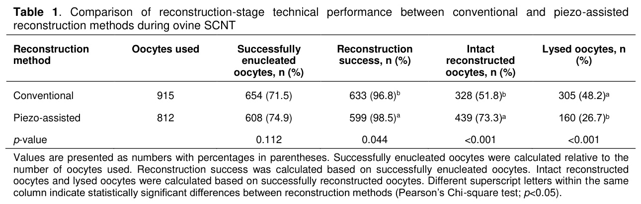
Values are presented as numbers with percentages in parentheses. Successfully enucleated oocytes were calculated relative to the number of oocytes used. Reconstruction success was calculated based on successfully enucleated oocytes. Intact reconstructed oocytes and lysed oocytes were calculated based on successfully reconstructed oocytes. Different superscript letters within the same column indicate statistically significant differences between reconstruction methods (Pearson’s Chi-square test; p<0.05).
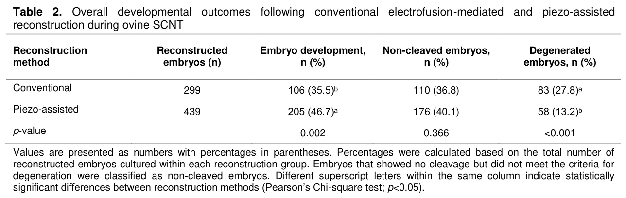
Values are presented as numbers with percentages in parentheses. Percentages were calculated based on the total number of reconstructed embryos cultured within each reconstruction group. Embryos that showed no cleavage but did not meet the criteria for degeneration were classified as non-cleaved embryos. Different superscript letters within the same column indicate statistically significant differences between reconstruction methods (Pearson’s Chi-square test; p<0.05).
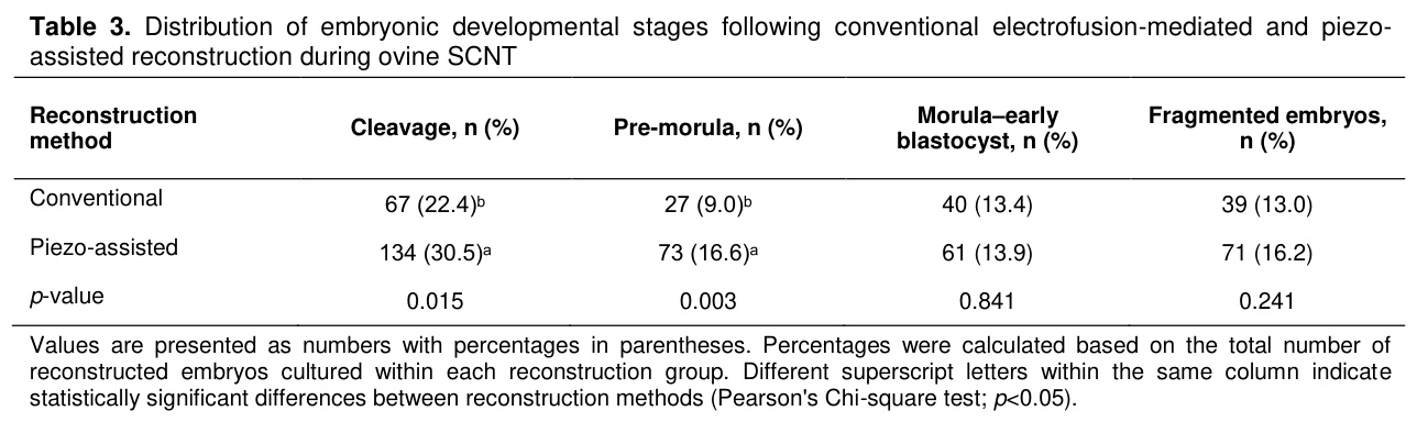
Values are presented as numbers with percentages in parentheses. Percentages were calculated based on the total number of reconstructed embryos cultured within each reconstruction group. Different superscript letters within the same column indicate statistically significant differences between reconstruction methods (Pearson's Chi-square test; p<0.05).
Tartışma
Somatic cell nuclear transfer is a multistep procedure in which each micromanipulation step influences the number of reconstructed embryos available for subsequent development. Therefore, optimization of reconstruction procedures remains essential for improving livestock cloning efficiency [14,15]. In the present study, conventional electrofusion-mediated and piezo-assisted reconstruction methods were compared during ovine SCNT. Piezo-assisted reconstruction significantly increased reconstruction success and the proportion of intact reconstructed oocytes, reduced oocyte lysis, and improved embryo development, cleavage, and pre-morula formation. These findings indicate that piezo-assisted reconstruction enhances post-manipulation oocyte integrity and supports early embryonic development. Because all recipient oocytes underwent an identical enucleation protocol, these differences can be attributed to the reconstruction procedure rather than enucleation itself. As reported previously, enucleation depends primarily on oocyte quality, accurate spindle identification, and operator proficiency, whereas reconstruction methods mainly influence post-manipulation oocyte integrity and subsequent embryo development [5,6,11,12,16,17].Although the absolute difference in reconstruction success between the two methods was modest, it was significantly higher in the piezo-assisted group (98.5% vs. 96.8%; p=0.044). Similar observations in ovine SCNT suggest that donor cell transfer is a reproducible technical step and that overall cloning efficiency depends on optimization of multiple procedures rather than a single micromanipulation technique [5,6,15,16,18]. In contrast, piezo-assisted reconstruction better preserved oolemma integrity by increasing the proportion of intact reconstructed oocytes and reducing oocyte lysis. Because membrane disruption immediately reduces the number of viable reconstructed embryos available for activation and subsequent development, preservation of membrane integrity is essential for successful SCNT. These findings agree with previous studies showing that piezo-assisted reconstruction minimizes manipulation-induced damage and improves membrane integrity [7,8,10,17,19], consistent with recent reviews highlighting the importance of reducing procedure-related cellular injury to improve SCNT outcomes [9,20].
The improved post-manipulation oocyte integrity observed after piezo-assisted reconstruction may be explained by controlled oolemma penetration using piezo-generated mechanical pulses. In this approach, permeabilized donor fibroblasts are deposited directly into the ooplasm without electrofusion, thereby minimizing membrane disruption and reducing oocyte lysis. Previous studies have similarly shown that less traumatic oocyte manipulation preserves membrane integrity, improves oocyte viability, and enhances reconstruction quality [7,10,11,17,19]. Collectively, these findings suggest that the principal advantage of piezo-assisted reconstruction is preservation of recipient oocyte integrity rather than a direct improvement in cloning efficiency [9,15,21,22].
Although embryonic development was evaluated as a secondary outcome, piezo-assisted reconstruction resulted in significantly higher rates of embryo development, cleavage, and pre-morula formation than the conventional approach, whereas morula–early blastocyst development remained comparable between the groups. These findings suggest that improved post-manipulation oocyte integrity primarily benefits early embryonic development by increasing the number of viable reconstructed oocytes available for activation and culture. However, this early developmental advantage did not translate into a higher rate of morula–early blastocyst development, suggesting that biological factors beyond the reconstruction procedure become increasingly important as embryogenesis progresses. Similar observations have been reported in ovine SCNT studies, where improvements in early embryo development did not necessarily translate into higher blastocyst production or cloning efficiency [18]. This limitation is likely attributable to incomplete donor nuclear reprogramming, defective embryonic genome activation, and inadequate epigenetic remodeling, which remain major barriers to successful cloning [23-26]. Overall, these findings indicate that refinement of reconstruction procedures improves early embryonic development but, by itself, is insufficient to overcome the biological limitations that continue to constrain cloning efficiency.
Sonuç
In conclusion, this study demonstrated that piezo-assisted reconstruction improves reconstruction-stage technical performance during ovine somatic cell nuclear transfer by modestly increasing reconstruction success, reducing oocyte lysis, and increasing the proportion of intact reconstructed oocytes. These improvements were associated with enhanced early embryonic development, particularly cleavage and pre-morula formation, whereas morula–early blastocyst development remained comparable between the two methods. Piezo-assisted reconstruction therefore represents a practical strategy for preserving oocyte integrity during SCNT. Further studies evaluating pregnancy establishment and live offspring production are needed to determine whether these advantages translate into improved cloning efficiency in sheep.References
- Wilmut I, Schnieke AE, McWhir J, Kind AJ, Campbell KH. Viable offspring derived from fetal and adult mammalian cells. Nature 1997; 385: 810-813.
- Wilmut I, Beaujean N, De Sousa PA, et al. Somatic cell nuclear transfer. Nature 2002; 419: 583-586.
- Wilmut I, Bai Y, Taylor J. Somatic cell nuclear transfer: Origins, the present position and future opportunities. Philos Trans R Soc Lond B Biol Sci 2015; 370: 20140366.
- Wang X, Qu J, Li J, et al. Epigenetic reprogramming during somatic cell nuclear transfer: Recent progress and future directions. Front Genet 2020; 11: 205.
- Birler S, Pabuccuoğlu S, Demir K, et al. Production of cloned lambs: Transfer of early cleavage-stage embryos to final recipients. Journal of the Faculty of Veterinary Medicine, İstanbul University 2010; 36: 1-8.
- Demir K, Pabuccuoğlu S, Cirit Ü, et al. Effects of serum starvation and ionomycin activation on the development of somatic cell nuclear transfer embryos in sheep. Journal of the Faculty of Veterinary Medicine, Ankara University 2019; 66: 37-42.
- Galli C, Lagutina I, Vassiliev I, Duchi R, Lazzari G. Comparison of microinjection (piezo-electric) and cell fusion for nuclear transfer success with different cell types in cattle. Cloning Stem Cells 2002; 4: 189-196.
- Lagutina I, Lazzari G, Duchi R, et al. Comparative aspects of somatic cell nuclear transfer with conventional and zona-free methods in cattle, horse, pig, and sheep. Theriogenology 2007; 67: 90-98.
- Czernik M, Anzalone DA, Palazzese L, Oikawa M, Loi P. Somatic cell nuclear transfer: Failures, successes and the challenges ahead. Int J Dev Biol 2019; 63: 123-130.
- Yang Y, Ding C, Wang E, et al. Piezo-assisted nuclear transfer affects cloning efficiency and may cause apoptosis. Reproduction 2007; 133: 947-954.
- Vázquez-Avendaño JR, Ambríz-García DA, Cortez-Romero C, Trejo-Córdova A, del Carmen Navarro-Maldonado M. Current state of the efficiency of sheep embryo production through somatic cell nuclear transfer. Small Rumin Res 2022; 212: 106702.
- Vázquez-Avendaño JR, Cortez-Romero C, Bravo-Vinaja Á, et al. Reproduction of sheep through nuclear transfer of somatic cells: A bibliometric approach. Animals (Basel) 2023; 13: 1839.
- Yagcioglu S, Ersoy N, Demir K, Birler S, Pabuccuoglu S. Can roscovitine and trichostatin A be alternatives to standard protocols for cell cycle synchronization of ovine adult and foetal fibroblast cells? Reprod Domest Anim 2023; 58: 1251-1260.
- Srirattana K, Kaneda M, Parnpai R. Strategies to improve the efficiency of somatic cell nuclear transfer. Int J Mol Sci 2022; 23: 1969.
- Malin K, Witkowska-Piłaszewicz O, Papis K. The many problems of somatic cell nuclear transfer in reproductive cloning of mammals. Theriogenology 2022; 189: 246-254.
- Campbell KHS, Fisher P, Chen WC, et al. Somatic cell nuclear transfer: Past, present and future perspectives. Theriogenology 2007; 68: 214-231.
- Iuso D, Czernik M, Zacchini F, Ptak G, Loi P. A simplified approach for oocyte enucleation in mammalian cloning. Cell Reprogram 2013; 15: 490-494.
- Hosseini SM, Moulavi F, Foruzanfar M, et al. Effect of donor cell type and gender on the efficiency of in vitro sheep somatic cell cloning. Small Rumin Res 2008; 78: 162-168.
- Kim EY, Park MJ, Park HY, et al. Improved cloning efficiency and developmental potential in bovine somatic cell nuclear transfer with the Oosight imaging system. Cell Reprogram 2012; 14: 305-311.
- Mrowiec P, Bugno-Poniewierska M. Technical, biological and molecular aspects of somatic cell nuclear transfer: A review. Ann Anim Sci 2022; 22: 63-87.
- Ma L, Liu X, Wang F, et al. Different donor cell culture methods can influence the developmental ability of cloned sheep embryos. PLoS One 2015; 10: e0135344.
- Gouveia C, Huyser C, Egli D, Pepper MS. Lessons learned from somatic cell nuclear transfer. Int J Mol Sci 2020; 21: 2314.
- Matoba S, Liu Y, Lu F, et al. Embryonic development following somatic cell nuclear transfer impeded by persisting histone methylation. Cell 2014; 159: 884-895.
- Liu W, Liu X, Wang C, et al. Identification of key factors conquering developmental arrest of somatic cell cloned embryos by combining embryo biopsy and single-cell sequencing. Cell Discov 2016; 2: 16010.
- Matoba S, Zhang Y. Somatic cell nuclear transfer reprogramming: Mechanisms and applications. Cell Stem Cell 2018; 23: 471-485.
- Li Y, Sun S, Xu Y, et al. Efficient somatic cell nuclear transfer by overcoming both pre- and post-implantation epigenetic barriers. Adv Sci (Weinh) 2025; 12: e04669.
- KOYUN KARACİĞER DOKU ARGİNAZININ GUANİDİN VE BAZI AMİNO ASİTLER TARAFINDAN İNHİBİSYONU VE İNHİBİSYONUN KİNETİĞİ
- KOYUNLARDA ÜRİNER SİSTEMİN NORMAL VE HASTALIKLI YAPILARININ ULTRASONOGRAFİK İNCELENMESİ
- ELAZIĞ BÖLGESİ KOYUNLARINDA GÖRÜLEN AYAK HASTALIKLARININ KLİNİK OLARAK DEĞERLENDİRİLMESİ
- KOYUN VE KEÇİLERDE BÖBREK HİDATİDOZİSİNİN İNSİDENS VE PATOLOJİSİ
- ELAZIĞ YÖRESİNDEKİ SÜT İNEĞİ VE KOYUN BESLENME PROGRAMLARININ DÖL VERİMİNE ETKİLERİ *